

White-Matter-Nulled 3D MPRAGE
Unlock new contrast and reveal more across the central nervous system
Hear from the clinicians
Expert Testimonials: Neurologists, Neurosurgeons, Neuroradiologists
Leading clinicians share how White Matter Nulled 3D MPRAGE has changed their practice:
- Neuroradiologist: spinal cord lesion detection
- Neurologist: longitudinal follow-up and treatment monitoring in multiple sclerosis
- Neurosurgeon: better surgical planning and confidence for intramedullary lesions or syringomyelia
Clinical Applications – Complete CNS Coverage
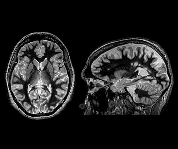
Brain
- Deep Brain Stimulation planning
Superior visualization of thalamic subnuclei for DBS target delineation. - Brain tumors
Cortical infiltration assessment and tumor margin delineation. - Multiple sclerosis
Detection of cortical, juxtacortical and thalamic lesions. Neurodegenerative disease
Cortical and subcortical grey matter evaluation and related conditions.
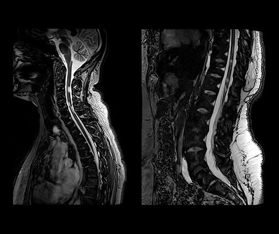
Spinal Cord
- Intramedullary lesions
Focal spinal cord signal abnormalities, improved conspicuity vs conventional T2 or STIR in difficult-to-image segments - Multiple sclerosis
Cervical, cervicothoracic and thoracic lesion detection, same contrast principle as brain acquisition for consistent reader experience. Improved lesion burden assessment and longitudinal follow-up consistency, with up to 51% more lesions detected* - Motion and artifacts
3D acquisition with optimized parameters reduces sensitivity to common artifacts encountered with standard T2/STIR sequences - Thoracic spine
The thoracic cord remains one of the most challenging segments to image. WMn 3D MPRAGE provides improved lesion conspicuity vs STIR/T2 in this region, with 3D multiplanar capability.
Optimized for Clinical Practice
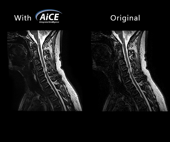
Fast3D + AiCE – Maximum Quality, Minimum Time
Combining Fast3D acceleration and Advanced intelligent Clear-IQ Engine (AiCE) deep learning reconstruction maximises image quality in WMn acquisitions. High SNR is maintained even at reduced scan times: making WMn 3D MPRAGE a practical, protocol-ready addition to any standard neurological MR exam.
- AiCE deep learning denoising preserves anatomical details
- Fast3D reduces acquisition time without SNR penalty combined with AiCE
- 1 mm isotropic resolution achievable in under 5 minutes for the spinal cord
- Compatible with 3T and 1.5T MR systems

“We believe this new 3D White-Matter-Nulled MPRAGE approach represents a real paradigm shift in spinal cord imaging compared to traditional Sagittal T2 and STIR, with strong potential impact on patient management.”
Prof. Thomas Tourdias
Neuroradiologist
Head of the Neuroimaging Department
Bordeaux University Hospital, France

“The advantage of using White-Matter-Nulled MPRAGE sequence is that multiple sclerosis lesions could appear more visible against the suppressed white matter background, facilitating the identification of subtle lesions that might otherwise remain undetected.”
Prof. Aurélie Ruet
Neurologist
Head of the Department of Neurology and Inflammatory Diseases of the CNS
Bordeaux University Hospital, France

“Such an MRI sequence will guide the diagnosis and the surgery planning for a biopsy or a resection. In the near future, better definition between the tumor and the gray matter organization might identify safer entry zones.”
Dr. Paul Roblot
Neurosurgeon
Bordeaux University Hospital, France
KEY BENEFITS
Why White-Matter-Nulled 3D MPRAGE changes the diagnostic picture?




References
- Munsch F, Ravache A, Yamamoto T, et al. Enhanced Spinal Cord Lesion Detection in MS Using White-Matter-Nulled 3D MPRAGE with Deep Learning Reconstruction. AJNR Am J Neuroradiol. 2026;47(1):225-233. Published 2026 Jan 5. doi:10.3174/ajnr.A8950
- Roblot P, Vignes JR, Tourdias T. Images in neuroscience: central canal kinking in Chiari type I with syringomyelia. J Clin Neurosci. 2026;149:112008. doi:10.1016/j.jocn.2026.112008
- Planche V, Su JH, Mournet S, et al. White-matter-nulled MPRAGE at 7T reveals thalamic lesions and atrophy of specific thalamic nuclei in multiple sclerosis. Mult Scler. 2020;26(8):987-992. doi:10.1177/1352458519828297
- Tourdias T, Saranathan M, Levesque IR, Su J, Rutt BK. Visualization of intra-thalamic nuclei with optimized white-matter-nulled MPRAGE at 7T. Neuroimage. 2014;84:534-545. doi:10.1016/j.neuroimage.2013.08.069
- Saranathan M, Tourdias T, Bayram E, Ghanouni P, Rutt BK. Optimization of white-matter-nulled magnetization prepared rapid gradient echo (MP-RAGE) imaging. Magn Reson Med. 2015;73(5):1786-1794. doi:10.1002/mrm.25298
- Fournet, A., Munsch, F., Koubiyr, I. et al. Interthalamic adhesion alterations in multiple sclerosis: associations with thalamic damage and cognition. J Neurol 273, 535 (2026). https://doi.org/10.1007/s00415-026-14069-5
Social Media


