We are sorry we can't show you this video – as matter of your Cookies preferences. Accept here the Targeting Cookies to watch the video content.
Wide Field Swept Source OCT in One Single Capture
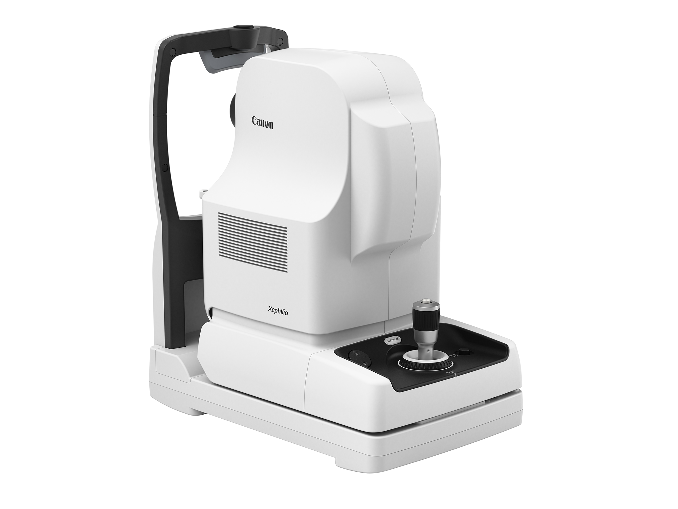
With the Xephilio OCT-S1 Canon Medical introduces revolutionary swept source technology allowing you to capture wide-field images of up to 23 mm in a single scan. The Xephilio OCT-S1 enables superior penetration of ocular opacities and provides outstanding tomographic images. Experience a new quality of OCT images in a single scan with greatly reduced noise, increased detail and improved visibility within just seconds. Swept source technology allows for a high scanning speed of 100,000 A-scans per second, while maximizing the essential signal data quantity during the acquisition. The 1,060 nm wavelength results in invisible scan lines and therefore ensures better patient collaboration and reduces the impact of the total examination.
Wide Field imaging
With Xephilio OCT-S1 wide-field images of up to 23 mm width can be acquired in just one single scan, equaling an incredible 80° viewing angle.
See the bigger picture
Canon OCT-S1 with 23 mm single capture vs a standard OCT. Canon Wide field OCT can visualise the a much larger area of retina not to miss any pathology in the periphery.
Single Capture Wide Field OCT Angiography
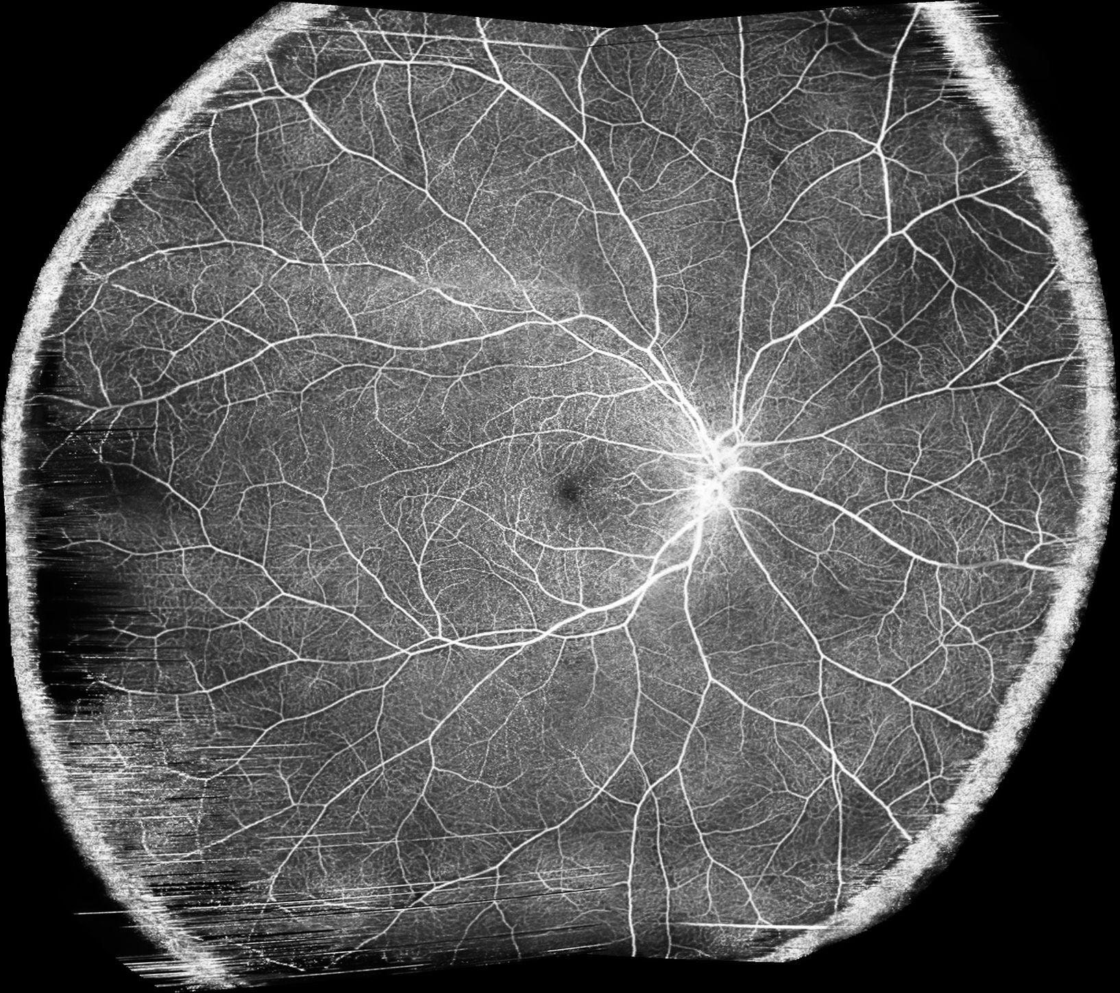
The Canon RX Capture software for OCT-S1 includes OCT Angiography already as standard! Wide field OCTA images of 23 x 20 mm can be obtained with a single acquisition. Visualization of Non – Perfusion areas over a very large surface is possible.
New AI Segmentation using Deep Learning
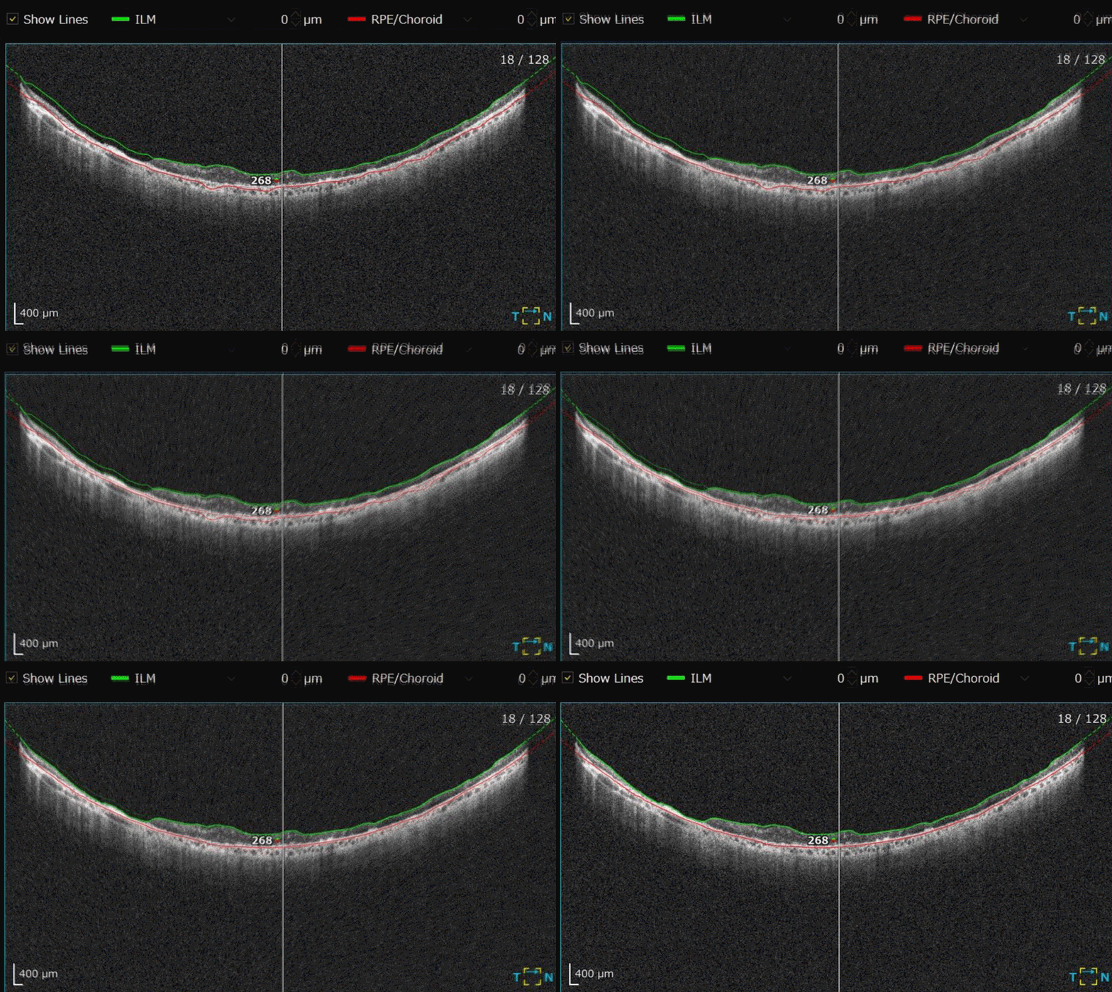
Thousands of tomograms, were manually segmented and used to generate the Training Data Set . The accuracy of determining the exact layer boundaries has been greatly improved and the number of detected layer boundaries is changed from 6 to 8.
Mosaic Wide Field OCTA
With the optional auto montage function, by using just 4 or 5 images, a mosaic image with a scan width up to ≈ 31 mm and a scan height up to ≈ 27 mm can be created. On the diagonal this combined image would have a length of approx. 36 mm or a 120° viewing angle.
5.3 mm depth of imaging
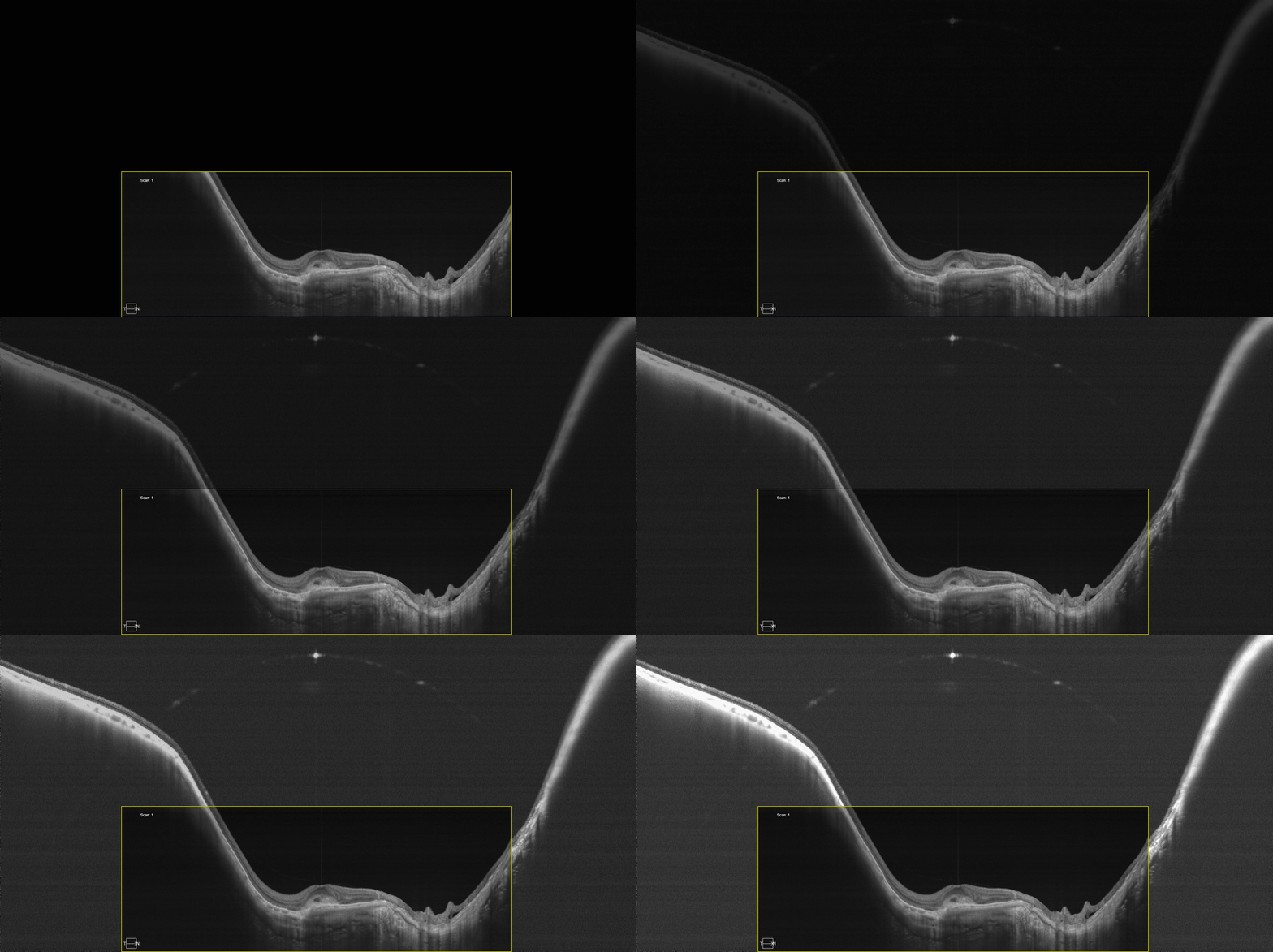
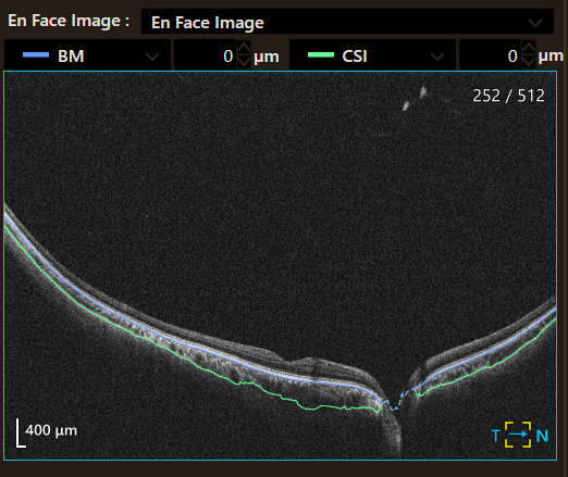
The 5.3 mm depth of imaging allows for detailed visualization of vitreous body and choroid in just a single scan with superior image quality
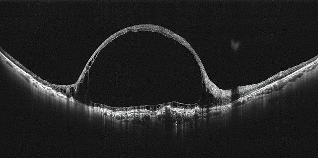
This 23mm wide angle scan nicely depicts a chronic central retinal vein occlusion with edema.
Courtesy of Dr. Kadomoto, Kyoto University, Japan
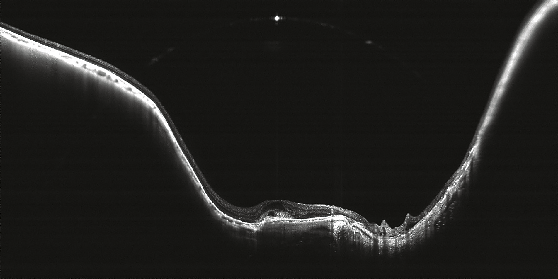
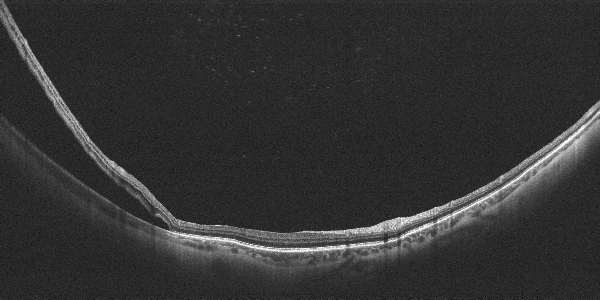
The curvature of the retina (especially posterior staphyloma) is well visualized in this Myopic Choroidal Neovascularization (mCNV) thanks to the 5.3 mm scan depth.
Courtesy of Dr. Kadomoto, Kyoto University, Japan
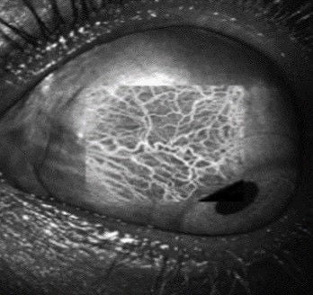
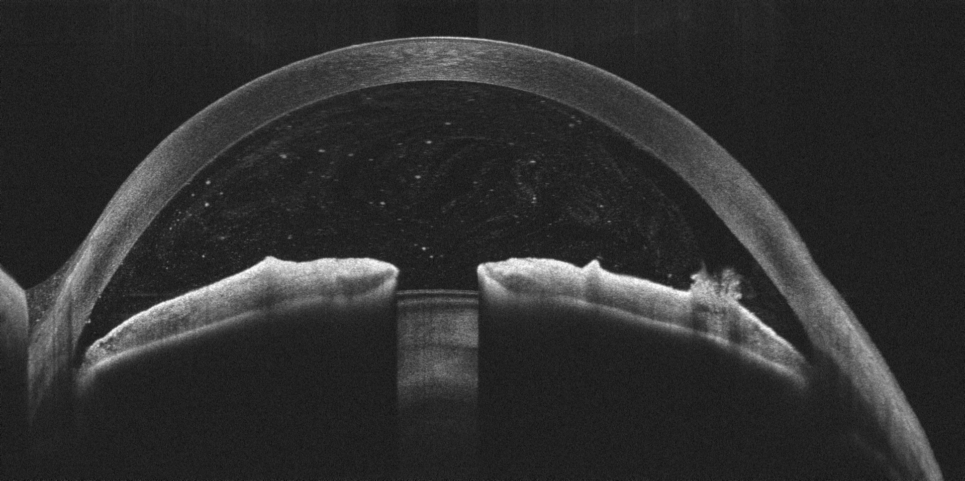
Anterior segment imaging
Easy and quick operation
Real-time tracking
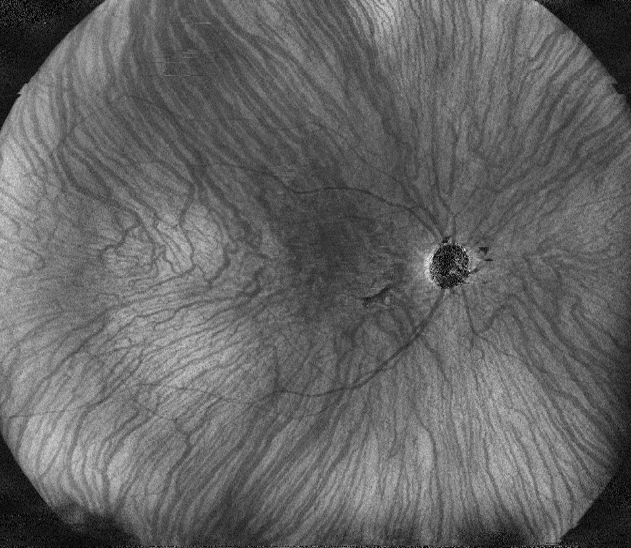
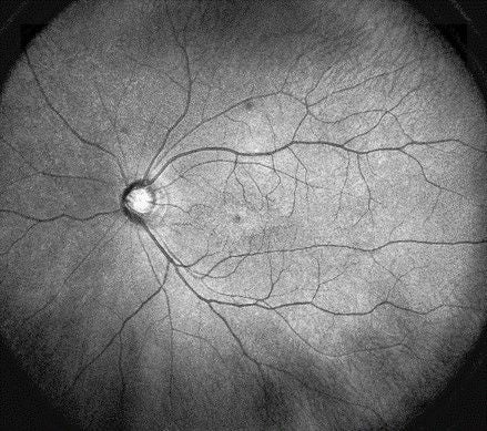
The built-in SLO provides real time retinal tracking during scanning. By detecting and compensating small involuntary eye movements, motion artefacts are greatly reduced resulting in a better image quality. The SLO technology also allows for accurate follow-ups, finding the same scan position with great precision. In addition the wide field SLO image gives a clear observation.
Pin-Point precision
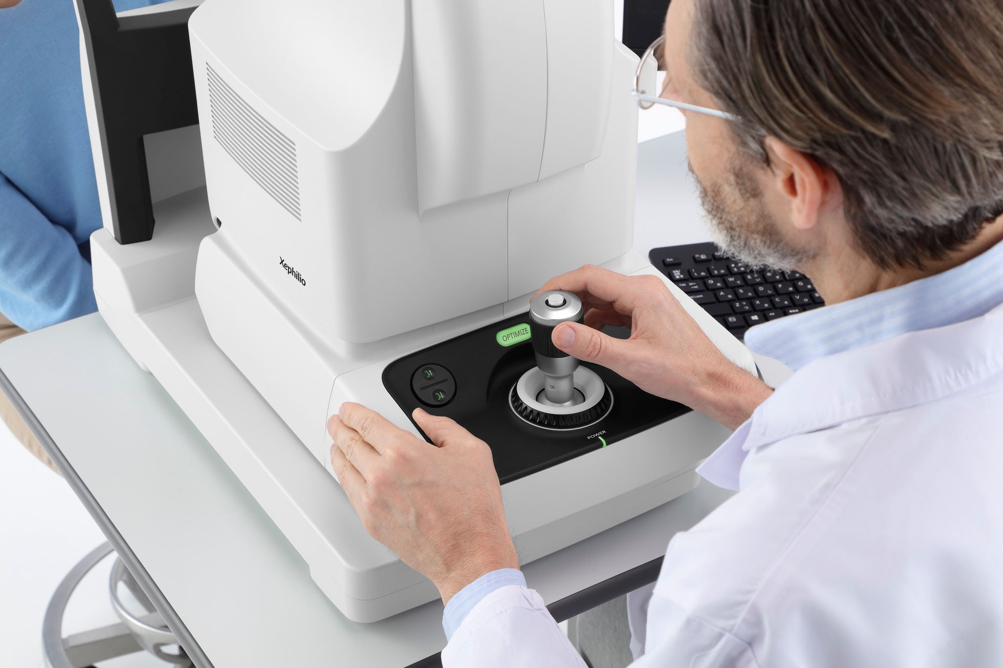
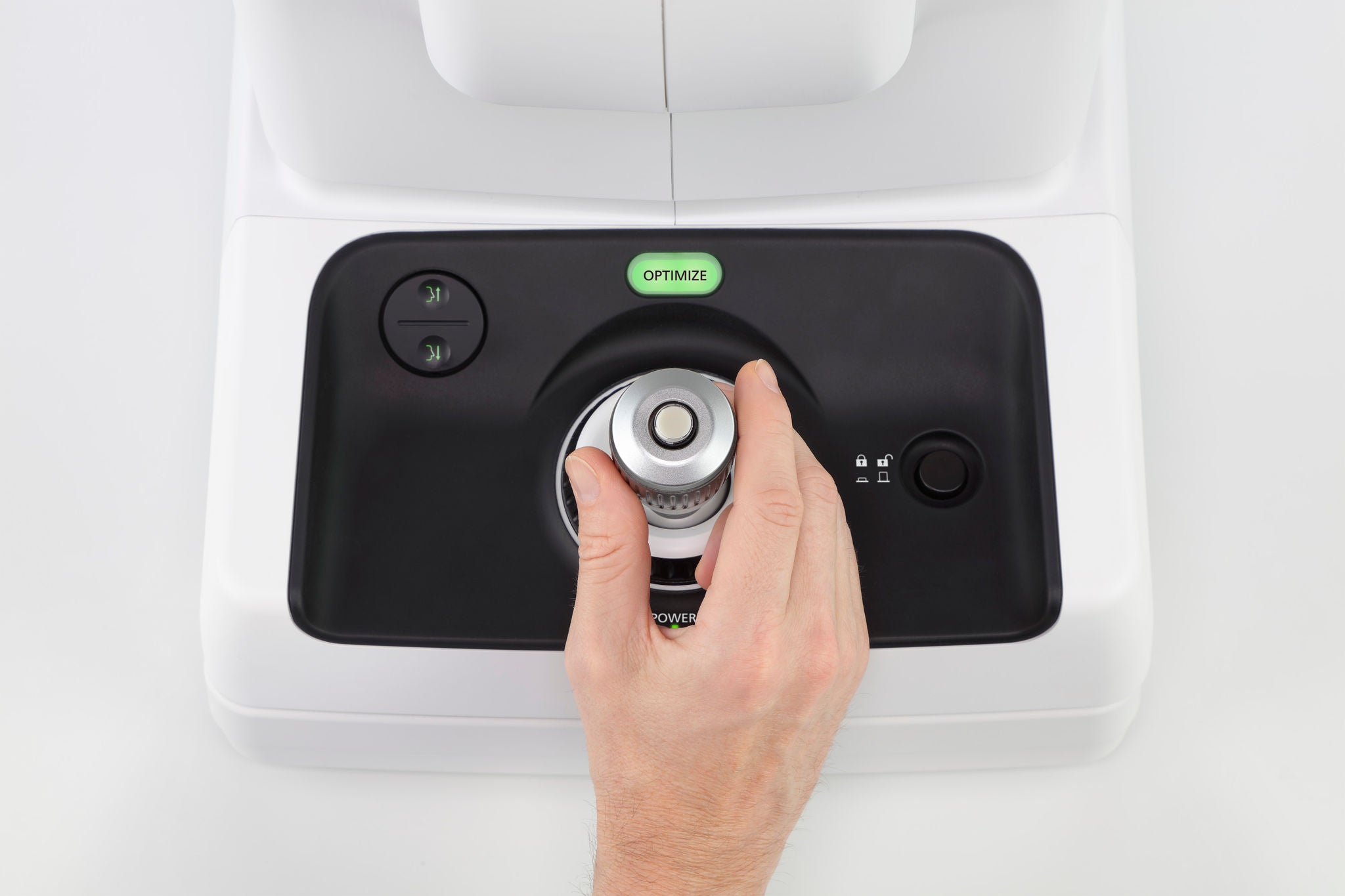
The OCT-S1 is equipped wih a joystick for easy, quick operation but combined with pin-point precision.
AI based optimize function
The optimize function enables several automated function: the fine alignment, auto focus, auto C-gate for optimal signal. The use of AI technology speeds up the adjustment enormously and also contributes to accuracy for best signal.
These functions combined with the SLO based real-time retinal tracking, make obtaining a good quality scan effortlessly!