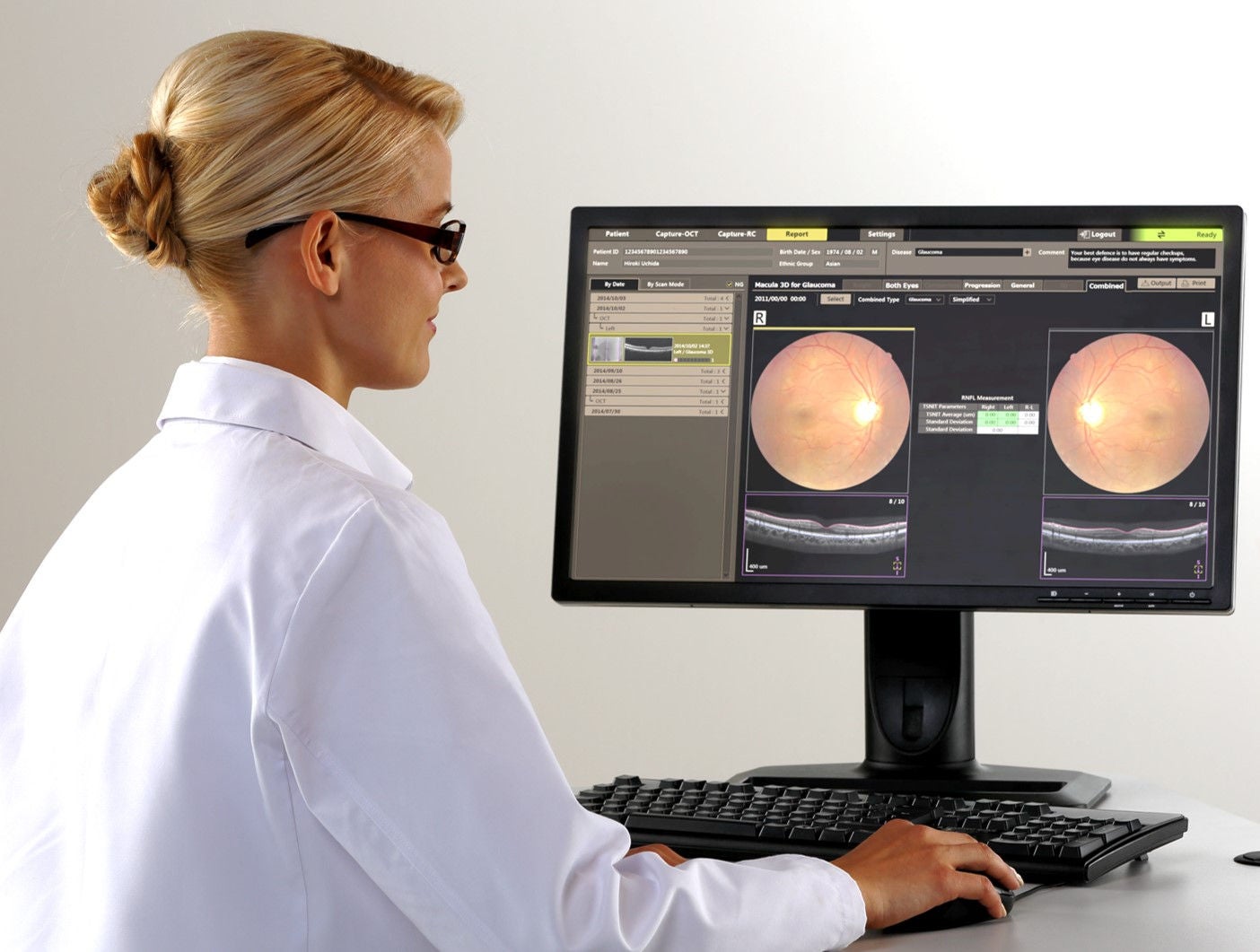
Canon’s Retinal Expert Capture Software allows for easy image acquisition and provides various functions for browsing, processing, printing, saving, and comparing images.
Perfect workflow
RX Capture software provides you with a perfect workflow. It is possible to start the RX Software directly from your EMR Software with a so-called command line interface. RX Software will automatically open on that patient, for image acquisition or viewing a report. Seamless integration for a perfect workflow.
Patient input
Alternatively the RX Software also can increase the efficiency of your workflow by importing a patient list directly from your EMR or by the use of a DICOM worklist. Of course patient information can also be input manually.
Cache functionality
RX software allows for much faster to previous studies, as required for follow up and progression by the cache function. This functionality reduces waiting time for downloading images due to limited network speed considerably.
Fast, consistent exams - high patient comfort
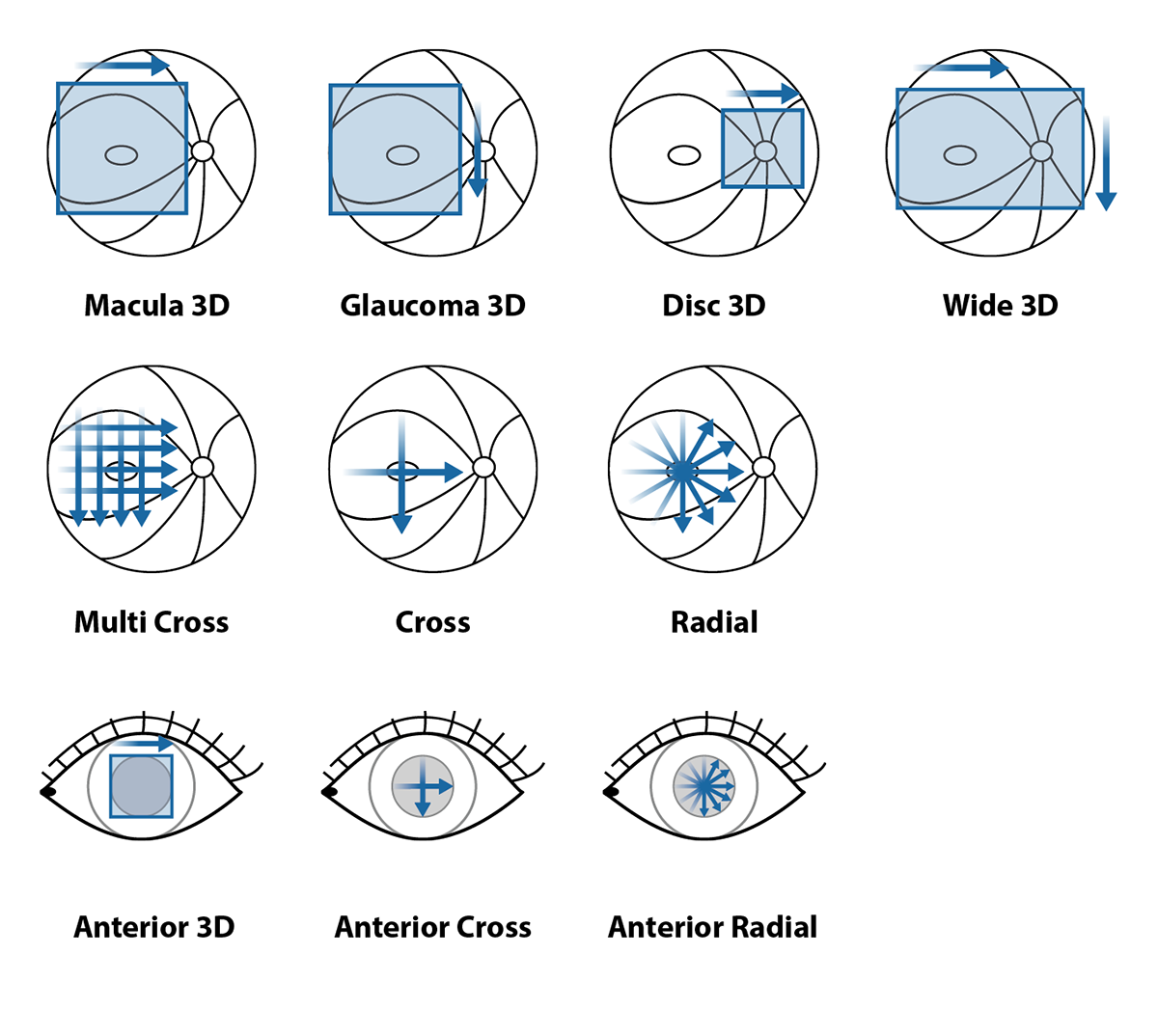
Xephilio OCT-A1 offers 10 fixed and freely programmable examination sets, allowing you to combine multiple scan modes into a single exam. Using examination presets can help you improve the workflow and consistency of exams especially when delegating.
Macula Examination
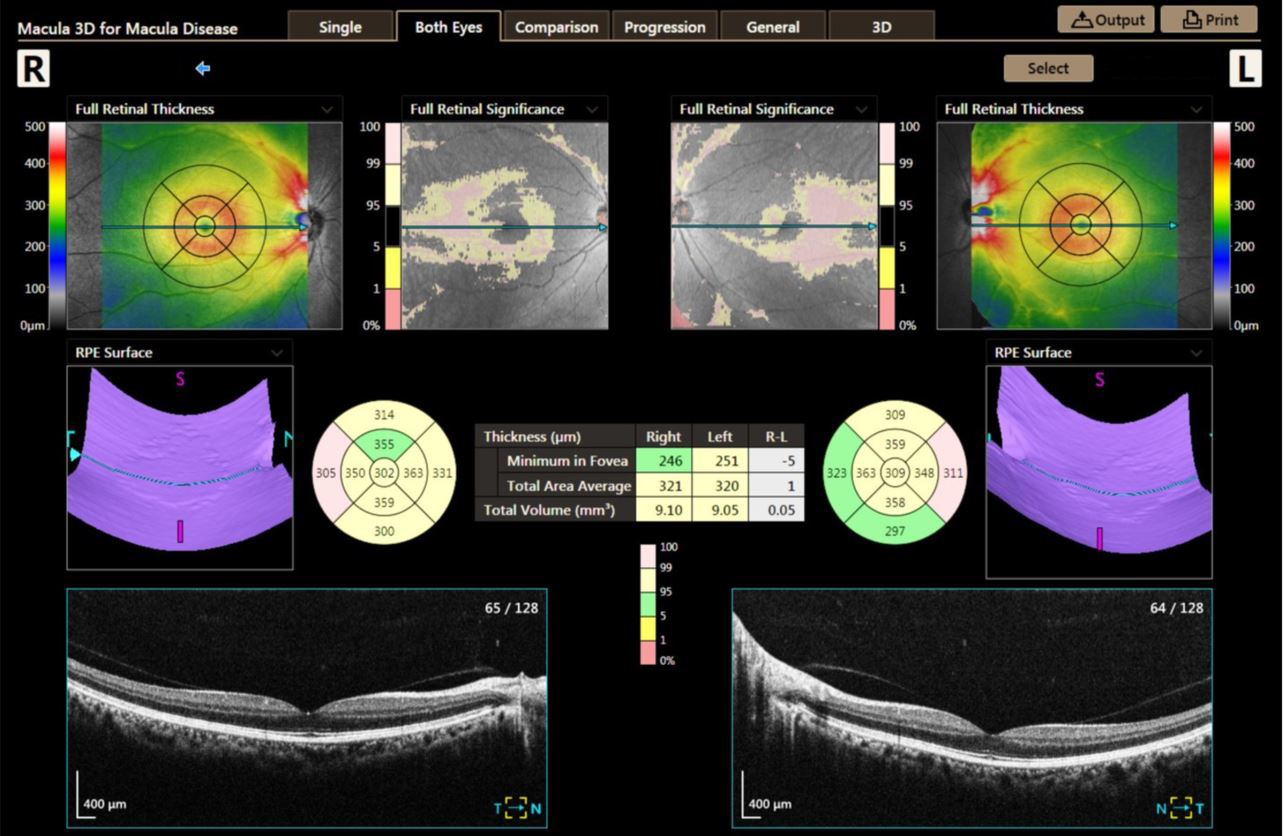
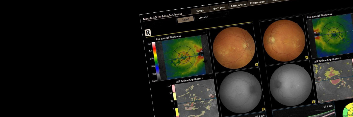
The system provides a detailed analysis of retinal thickness using comparisons with a normative database, ETDRS grids, various tables and 3D visualizations.
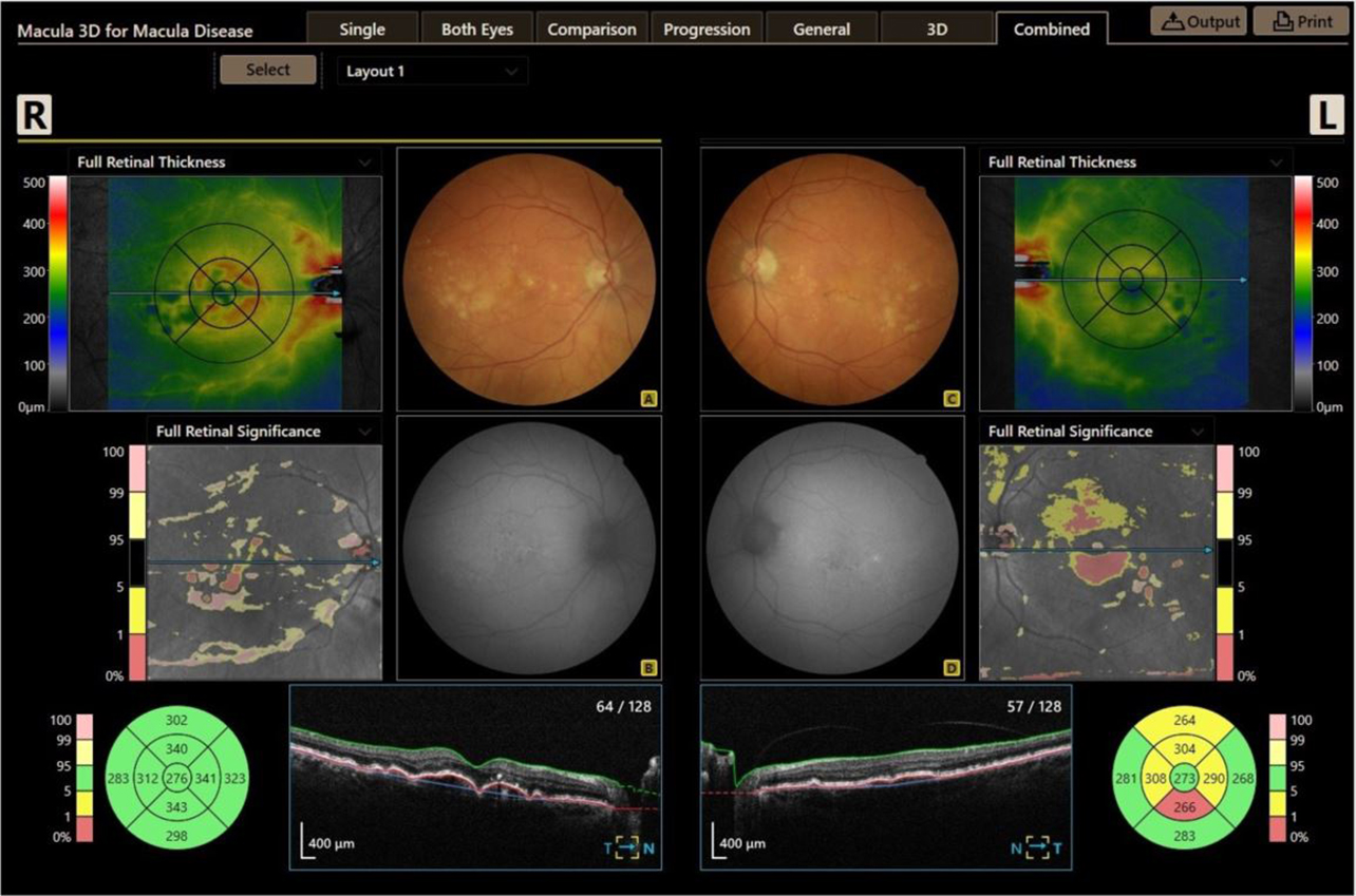
COMBINED REPORT
By importing fundus images from a retinal camera they can be easily integrated into the OCT evaluation. Fundus and OCT images can be displayed side by side or mapped and superimposed as needed. When using a Canon Retinal camera, the patient database can even be shared for a most efficient workflow.
Glaucoma Examination
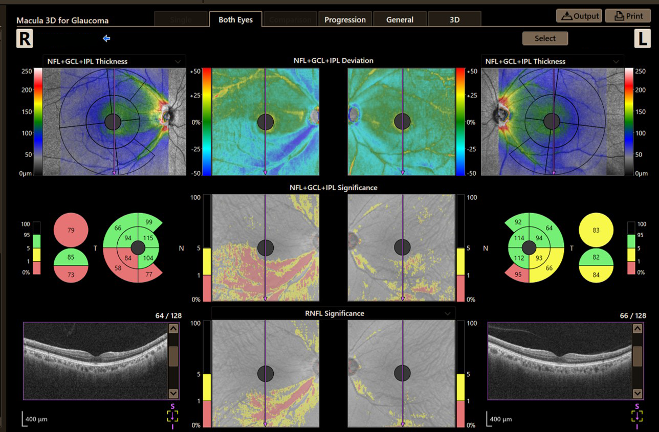
Macula 3D for glaucoma
Early detection is the key to slowing the progression of glaucoma. Xephilio OCT-A1 supports NFL + GCL + IPL and GCL + IPL measurements with a wide set of graphical representations for complete analysis.
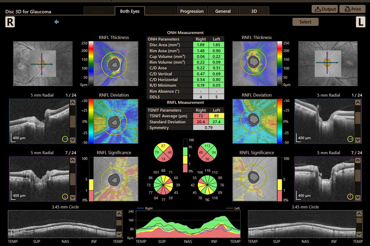
Disc 3D
Xephilio OCT-A1 allows a comprehensive analysis of all optic disc parameters, including comparisons with an extensive normative database.
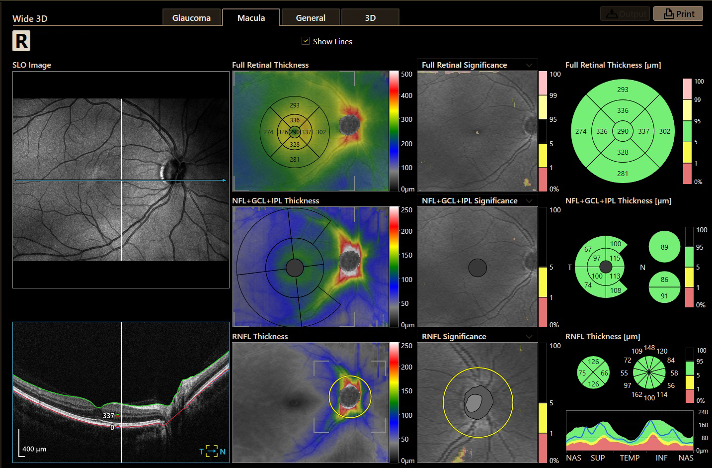
Wide 3D scan mode
This Wide 3D scan mode (13x10 mm) enables to perform a very efficient and time saving Glaucoma scan, determining not only the thickness of the NFL+GCL+IPL, but also the RNFL thickness around the optic disc: all in just one scan!! Results can be shown in the new combined Glaucoma 3D report.
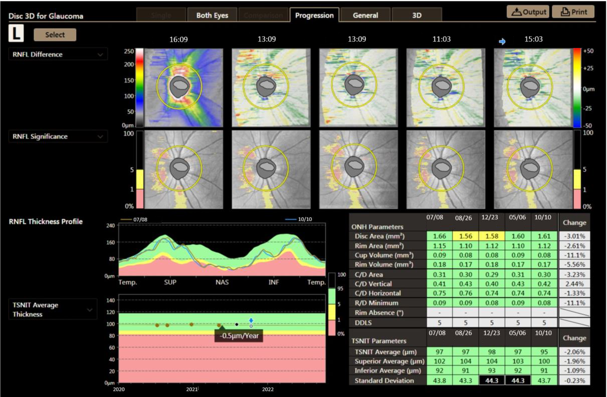
Progression
Analysis results comparing five examinations can be displayed to monitor progression.
Anterior Segment analysis
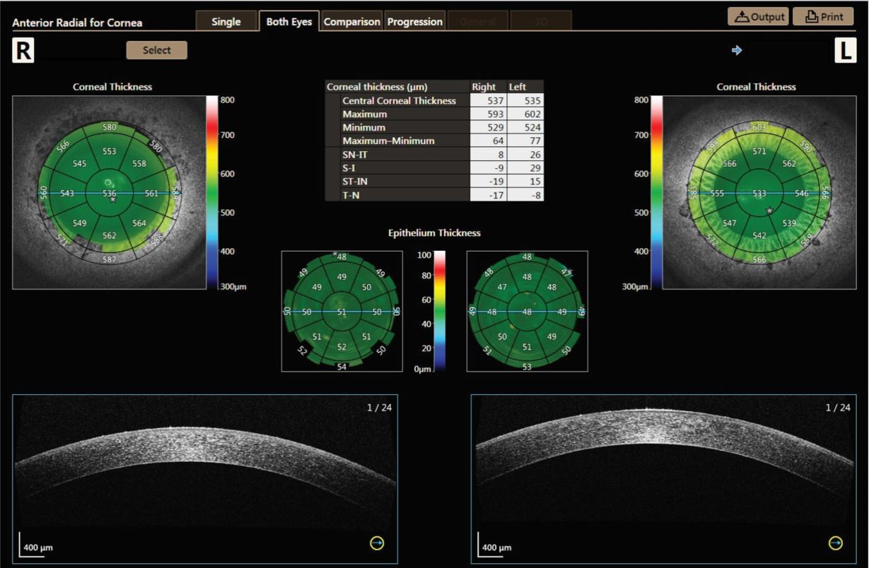
With the optional Anterior Segment Adapter ASA-1, Xephilio OCT-A1 also gives you the ability to analyze and document the anterior segment of the eye. The measurement tools allows to quantify standard parameters quickly and easily. Report are available for Single Eye, Both Eyes, Comparison and Progression.
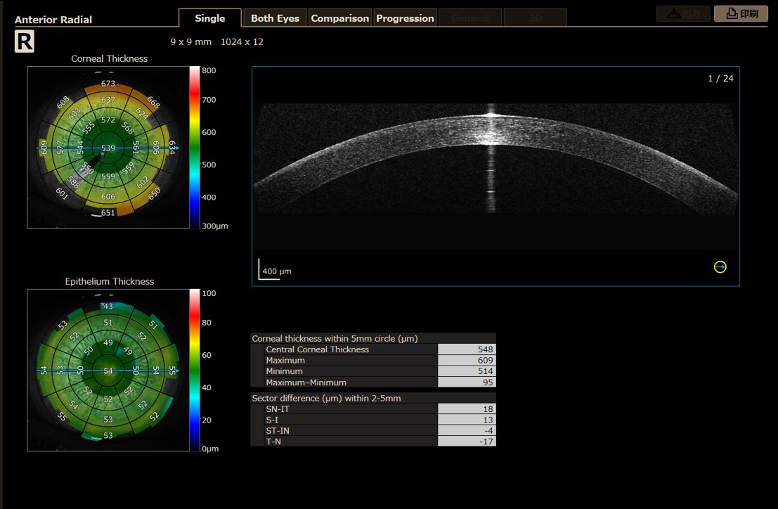
The new 9 mm wide anterior radial scan has a report layout as before, but offers a new wider analysis grid.
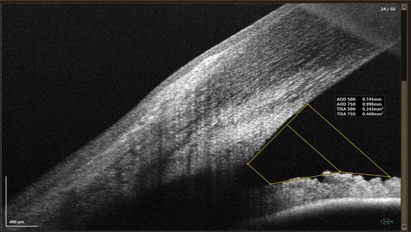
It also possible to analyze the distance between 2 points, angles and AOD (Angle Opening Distance) and TISA (Trabecular Iris Space Area).