

Clinical Applications of High-Resolution Imaging in the Abdomen
Katsuyoshi Ito, MD, PhD
This report discusses the clinical applications of various technologies for imaging of the abdomen, including Advanced intelligent Clear-IQ Engine (AiCE) and Precise IQ Engine (PIQE), which are used for high-precision T2-FSE imaging, Modified Fast 3D mode, which is used for high-precision dynamic imaging, and Expanded SPEEDER (Exsper), which is used for small-FOV high-resolution DWI imaging. AiCE and PIQE are Deep Learning Reconstruction (DLR) technologies developed by Canon Medical Systems Corporation.
High-precision T2-FSE imaging with AiCE and PIQE
1. Overview of PIQE in MRI
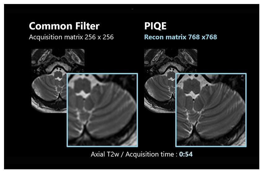
PIQE is a new technology which is based on Deep Learning. It allows low-SNR images, low-resolution images to be reconstructed to obtain high-SNR images, high-resolution images. When applied to MRI, PIQE employs two DLR technologies: denoising and up-sampling. Zero-fill interpolation is performed during processing, but the limitations of this approach are that some image blurring cannot be eliminated and ringing artifacts are generated. To overcome these limitations of zero filling, the neural networks in PIQE are trained with high-resolution images acquired by full sampling used as the teaching data.
2. High-precision T2-FSE with PIQE
The quality of T2-weighted fast spin echo (T2-FSE) images can be improved by employing PIQE.
Figure 1 shows an enlarged view of the pancreas. The spatial resolution is increased by a factor of 2 to 2.5 when PIQE is applied (b), reducing blurring and more clearly delineating the margins of the pancreas, the pancreatic duct, and other structures in the abdominal cavity. Such high-precision images can also be obtained in scans with fat suppression used in combination.
Figure 2 shows images of a patient with a hepatocellular carcinoma. The margins of the tumor and the linear hyperintensities within the tumor are more clearly visualized when PIQE is applied (b). In addition, an aortic dissection flap and the internal characteristics of the tumor in the right adrenal gland are demonstrated with outstanding clarity.
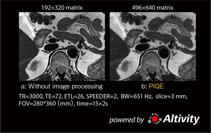
Figure 1: Effects of PIQE: high-precision T2-FSE imaging
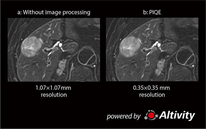
Figure 2: Fat-suppressed T2-FSE in a patient with hepatocellular carcinoma
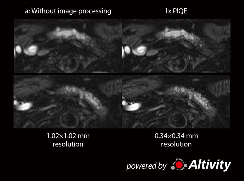
High-precision dynamic imaging in Modified Fast 3D mode
1. Modified Fast 3D mode
It is possible to perform high-precision dynamic imaging by using the fast-scanning technique known as Modified Fast 3D mode in combination with AiCE.
Modified Fast 3D mode is a scanning technique which includes modifications to conventional Fast 3D mode, in which data is acquired in a wheel-shaped pattern. In Modified Fast 3D mode, the asymmetric half Fourier method (AFI) is used in combination to sequentially acquire data in both the slice- and phase-encoding directions. Scan times are reduced by performing skipped data acquisition in a bow-shaped pattern. In addition, the k-space is mainly filled with stable signals by filling the data sequentially, which reduces blurring and improves image quality. Another advantage of Modified Fast 3D mode is that image blurring is less of an issue when the Fast 3D factor is increased.
Even when the scan time is reduced by approximately 25% compared to conventional imaging methods, the same level of image quality is maintained. Therefore, high-precision dynamic imaging is ensured because the scan time can be shortened while maintaining high resolution. Compared to other fast-scan techniques such as Compressed SPEEDER (CS), image blurring of the entire image is minimized, allowing the margins of the intrahepatic blood vessels and other structures to be more clearly visualized.
At our hospital, we previously performed dynamic imaging using SPEEDER or CS combined with AiCE, but we found that these approaches suffered from a number of limitations. With SPEEDER, we would see linear artifacts in the liver during the arterial phase and blurring of the entire image at higher AiCE levels, and with CS, we would observe overall image blurring and artifacts which appeared as white edges around structures. We have been able to overcome these limitations by employing Modified Fast 3D mode combined with AiCE. This approach minimizes image blurring and artifacts, allowing us to obtain high-quality images in which the margins of structures are accurately depicted.
2. High-precision dynamic imaging
Figure 4 shows dynamic imaging of a patient with fatty liver performed using Modified Fast 3D mode (a). The peripheral branches of blood vessels are clearly seen even in the arterial phase. In the hepatocellular phase, the intrahepatic blood vessels are more clearly depicted in the Modified Fast 3D mode image (c) than in the CS image (b).
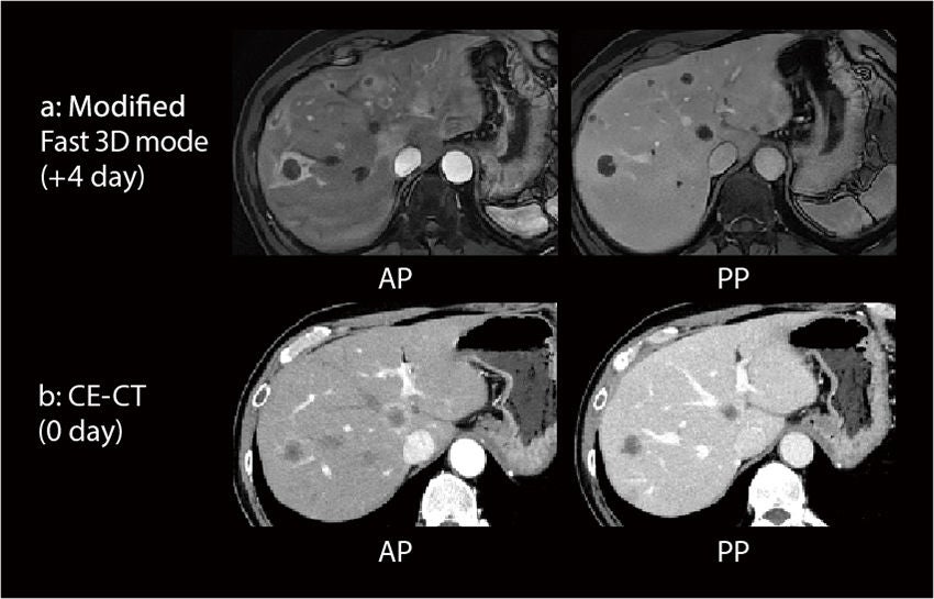
Figure 5 shows dynamic imaging of a patient with a hepatic hemangioma. In Modified Fast 3D mode images acquired in the arterial phase, a spotty hyperintensity in the lesion (a, red arrow) and multiple hyperperfusional abnormalities in the liver are clearly seen. In the hepatocellular phase, the intrahepatic blood vessels are more clearly depicted in the Modified Fast 3D mode image (c) than in the CS image (b).
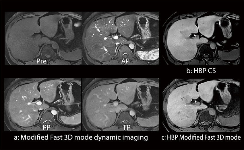
Figure 4: Dynamic imaging of fatty liver
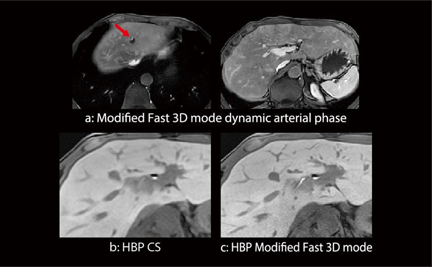
Figure 5: Dynamic imaging of hepatic hemangioma
Exsper DWI
1. Features of Exsper
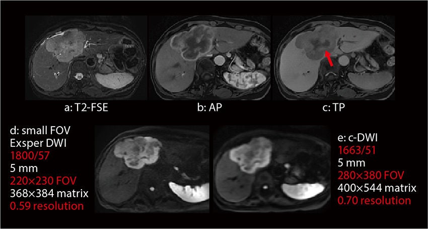
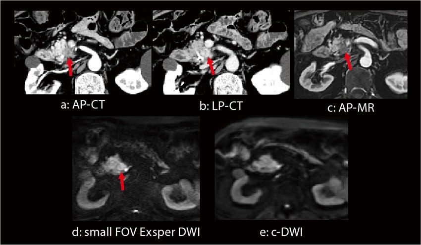
In Exsper, calibration data acquired during actual scanning is used to perform unfolding processing. Compared to SPEEDER, Exsper can reduce the artifacts arising from unfolding errors by calculating the weighting for unfolding in k-space. Artifacts near the center of the image caused by unfolding errors are less likely to occur when a small FOV is scanned, which helps to ensure high-resolution imaging.
3. Usefulness of breath-hold DWI
We conducted investigations to evaluate the quality of images acquired during free breathing and with breath-holding in clinical scans performed at our hospital. The lesion visualization score was found to be slightly higher for scanning during free breathing, but the difference was not statistically significant. However, breath-hold scanning was found to provide superior image quality in patients with lesions located under the diaphragmatic dome or near the edges of the liver, which tend to be affected by motion of the diaphragm or nearby segments of the intestinal tract as well as magnetic susceptibility artifacts. In such patients, breathhold scanning was judged to be clinically useful, with breathhold DWI considered to be most helpful when performed in addition to conventional DWI because breath-hold DWI allows scanning to be performed in a shorter time.//

Katsuyoshi Ito, MD, PhD
Professor, Department of Radiology,
Yamaguchi University Graduate School of Medicine, Japan
Professor, Department of Radiology,
Yamaguchi University Graduate School of Medicine, Japan
This article is a translation of the INNERVISION magazine, Vol.38, No.6, 2023.
Disclaimer
The contents of this report include the personal opinions of the author based on his clinical experience and knowledge.
Deep learning technology is used in the design stage of the image reconstruction processing for AiCE and PIQE. The system itself does not have self-learning capabilities.
Learn more about Canon Medical's MRI technologies and products

The content reflects information available at the time of publication and may differ from current information.
Social Media


