Search

Accessing Super High Resolution Without Compromise

Julien Savatovsky, MD
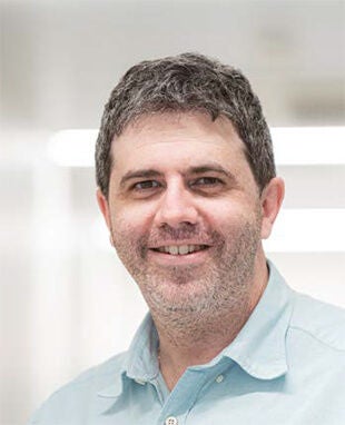
Dr Julien Savatovsky, a Neuroradiologist and the Deputy Head of Diagnostic Neuroradiology at Rothschild Foundation Hospital, Paris, France, uses advanced image reconstructions solutions, based on Artificial Intelligence (AI), in his daily routine. Collaborating closely with Canon Medical, he has contributed to the development of the latest reconstruction algorithms and evaluated them on the Hospital’s Vantage Orian 1.5T and Vantage Galan 3T MRI systems. This experience has convinced Dr. Savatovsky that AI can challenge the well-known ‘triangle of compromise’ in imaging between signal-to-noise ratio (SNR), spatial resolution, and acquisition time.
Image
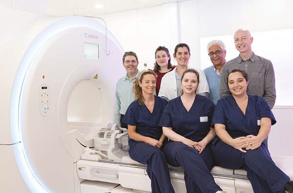
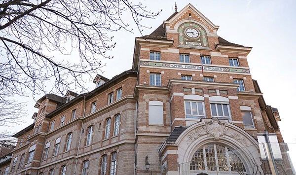
The Rothschild Foundation Hospital is a non-profit hospital in Paris, which pioneers care, research, and cooperation on eye and brain diseases. The Neuroradiology Department at the Hospital is continually involved in advancing research through the use of cutting-edge imaging techniques.
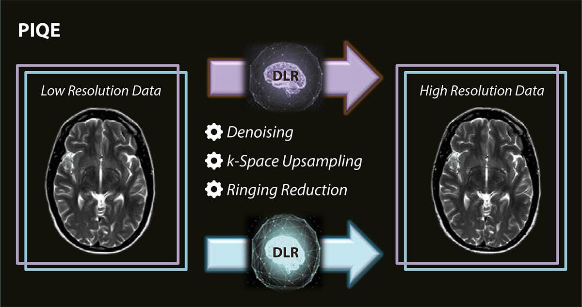
Canon’s Advanced intelligent Clear-IQ Engine (AiCE) was the first AI-based denoising solution made available for CT and MR imaging. It is a very robust solution that achieves high SNR images by selectively removing noise without affecting the signal coming from tissues.

Following the strong success of AiCE, Canon have recently released Precise IQ Engine (PIQE) - a new solution developed to increase image sharpness and gain in diagnostic confidence. Using several algorithms, PIQE removes noise, increases matrix size, and reduces Gibbs ringing artifacts. It produces sharp anatomical images that take MR imaging to the next level.

Image

‘Triangle of compromise’

“Image quality is crucial in diagnosis because it's what drives the confidence in the radiologist's diagnostics and the clinician’s confidence in the radiologist,” remarked Dr. Savatovsky.

“There are many subjective issues in the assessment of image quality and few objective measurements that we can do, including signal-to-noise ratio (SNR), spatial resolution contrast and contrast to noise ratio (CNR),” he continued. “Therein, lies what I call the ‘triangle of compromise’ that has ‘haunted’ MRI in that, for a given sequence you have a given time, a given SNR, and a given spatial resolution. If you adjust for better spatial resolution, for example, the consequences are two compromises - one on SNR that will go down and one other on the acquisition time that will be longer.”

Dr. Savatovsky has explored the impact of Deep Learning Reconstruction (DLR) techniques, such as Canon’s Advanced intelligent Clear-IQ Engine (AiCE) and the latest Precise IQ Engine (PIQE), in adressing these compromises.
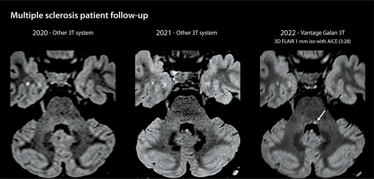
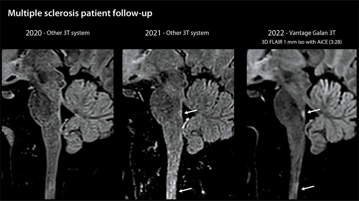
“Nowadays, Deep Learning technologies can be found in post-processing field, adding an extra layer to the reconstructed images, improving their quality", he said. "During years, we improved the SNR using filters, but it was at the cost of the image sharpness, due to the introduction of important blurring effects. And at the opposite, if we wanted a crisper image, we used high filter which also increase the noise. Now, thanks to Deep Learning recontructions such as AiCE, we have the possibility to improve SNR without compromising the sharpness of the images.”

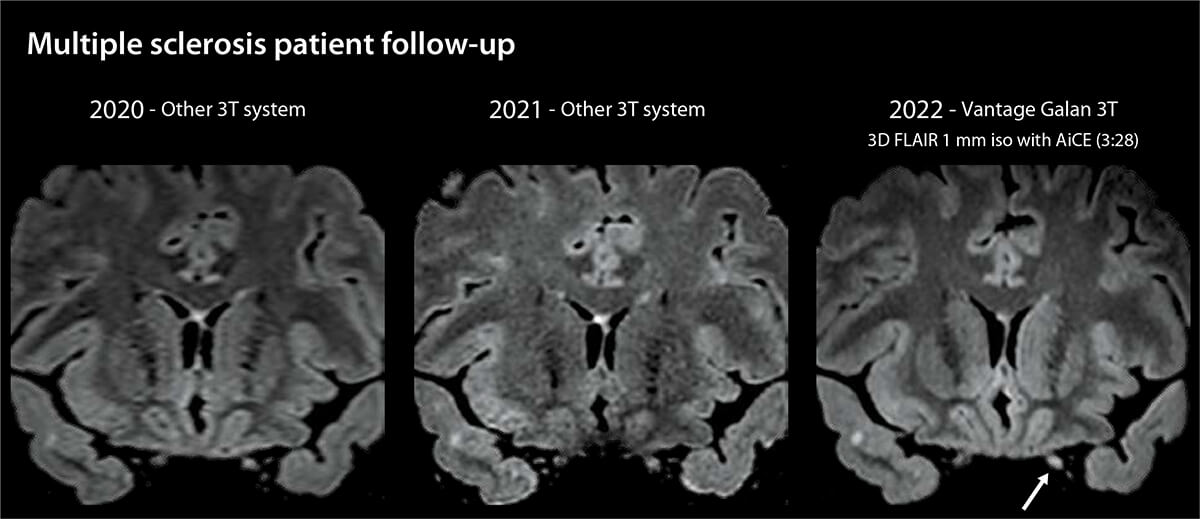
“In the below image, there is a small Multiple Sclerosis (MS) lesion of the posterior fossa that is very important to detect because it's usually enough to cause very significant symptoms,” explained Dr. Savatovsky. “The lesion is much more visible with AiCE technology thanks to contrast-to-noise ratio improvement.”
Image
“This cervical spinal cord lesion was not really visible previously. But with this one millimeter isotropic FLAIR reconstructed with AiCE, it was clearly visible without any doubts. By applying such reconstruction algorithm on 3D sequences, we get the benefits of the AI-based denoising in every planes," said Dr. Savatovsky.
Image
"The same patient had an optic neuritis. This is usually very challenging to observe it on a FLAIR sequence because of the low SNR that we get in this difficult region. Once again, AiCE allows the inflammation visualization despite the very high resolution, emphasizing the power of the algorithm," remarked Dr. Savatovsky.
Image
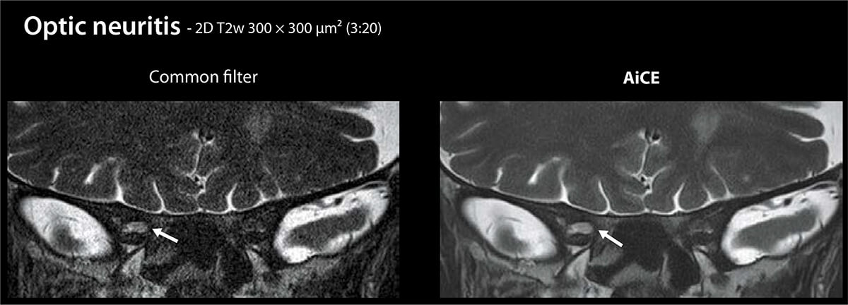
“This is an image created in three minutes with 300 Micron voxels on a different patient with a right optic neuritis. We want the voxel to be very small, but we end up with a very noisy image if we use common filters. When we apply Deep Learning reconstructions, we can see the optic neuritis really well and we recover some SNR and some CNR,” he added.
Image

Closing quality gaps

An other advantage of AiCE and PIQE is that they can help to close image quality gaps between systems, when a healthcare facility has different devices, such as 1.5T and 3T MRI systems.

“Thanks to the improvement in SNR, we can really enhance the image quality and get the same quality at 1.5T than the one we have at 3T,” said Dr. Savatovsky. “Just recently, I obtained an 800 micron isotropic image with a very high image quality at 1.5T, which we can usually only achieve on 3T systems.”
Dr Savatovsky and his team have performed a study to explore the image improvements allowed by DLR.

“We've proven on MS patients that both the SNR and the CNR were better with DLR. In addition, the stronger the DLR algorithm, the better the results,” he said. “Now, the ‘triangle of compromise’ is totally reconsidered. Using Deep Learning algorithms, we can improve the SNR, without compromise on spatial resolution and keeping the same scan time.”
Image

The power of PIQE

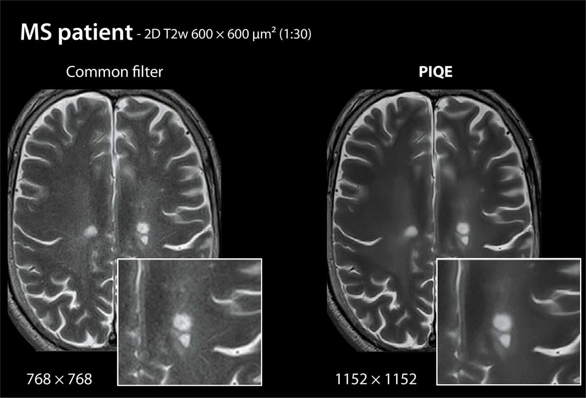
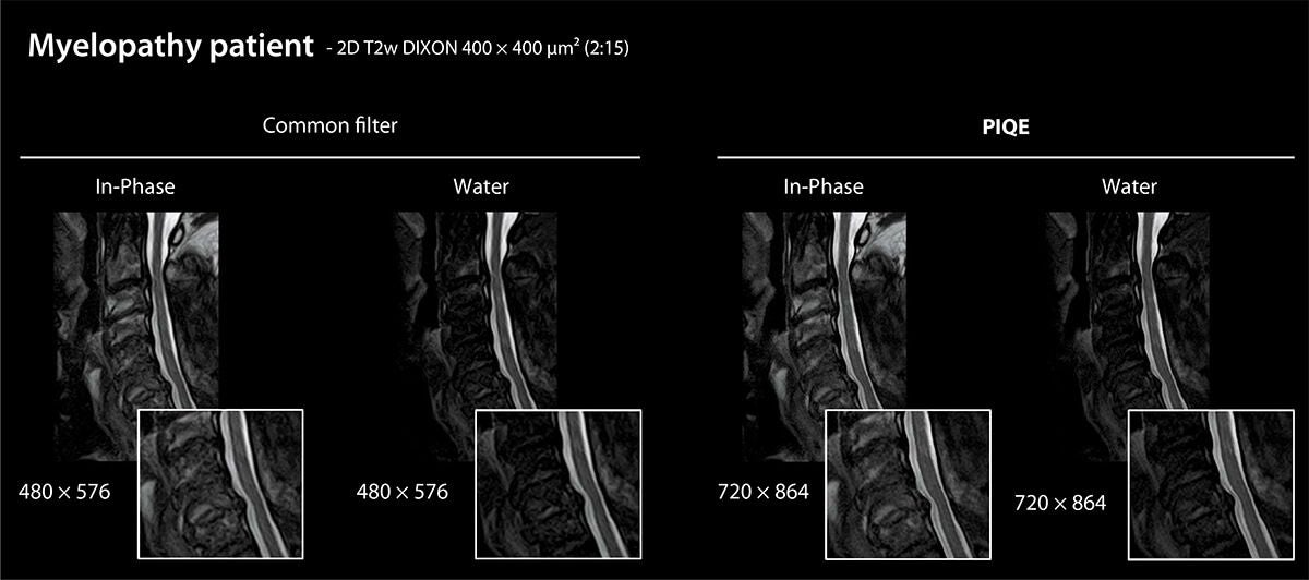
The team has also explored further by enhancing matrix resolution and image sharpness using PIQE.

“The first result that we've obtained using PIQE – which is currently only applicable on 2D MRI sequences – is that you can obtain sharper images without altering the SNR. We get now crisper images," said Dr Savatovsky.

“We feel that we get far more details, which is quite surprising, because it's the same acquisition and the same scan time. In addition of improving SNR, PIQE also increases the matrix resolution, facilitating contour depiction and helping us in difficult anatomy examinations. The structures are far crisper and we achieve a greater diagnostic confidence,” he continued.
Image

The future is here

With the experience already gained, Dr. Savatovsky believes some next steps in development might include application of AiCE and PIQE algorithms to more sequences.
“We are confident that Canon Medical is striving towards even further progress and we're really looking forward to this.” //
Image

“We're looking forward applying PIQE in 3D sequences and for low resolution sequences, such as diffusion imaging”

Julien Savatovsky, MD Deputy head of Diagnostic Neuroradiology at the Rothschild Foundation Hospital, Paris, France
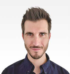
Dr. Julien Savatovsky
Dr. Savatovsky is Deputy Head of the Imaging Department (Diagnostic Neuroradiology) at the Rothschild Foundation Hospital, Paris, France.

Dr. Savatovsky continued his undergraduate training in medicine at the University of Paris-VI Pierre and Marie Curie and his internship in Paris. He completed his training with a two-year clinic in the Neuroradiology Department of the Pitié- Salpêtrière hospital group. He continued his hospital career as a hospital practitioner in neuroradiology at the Rothschild Foundation Hospital.

He specializes in nervous system imaging and neck diseases. In order to give access to high quality imaging to patients, Dr. Savatovsky uses specifically optimized 3T MRI equipment and has advanced training in MRI techniques (including sequence development and tuning) and in advanced post-processing.
Image
In addition to his clinical work and the management of the Imaging Department, Dr. Savatovsky has been involved in research for several years. He has contributed to over a hundred articles published in international journals.
His main areas of research include:
  • the development of new MRI sequences
  • imaging of the cerebral lymphatics and the blood-brain barrier
  • imaging of idiopathic intracranial hypertension
  • multiple sclerosis (MS) imaging, including high resolution sequences, magnetic susceptibility
  • innovative post-processing and AI.
He is a lecturer at various Parisian universities and at the European Course in Neuroradiology. He has also contributed to several radiology textbooks.
Image
Julien Savatovsky, MD
Deputy head of Diagnostic Neuroradiology at the Rothschild Foundation Hospital, Paris, France
Image
Valentin H. Prevost, PhD
MR Clinical Scientist
Canon Medical Systems Corporation
Testimonials, white papers, and articles about AI from users of Canon Medical Systems' AI technologies

Find more AI special articles

Real-World Application of AI

Read more
Image

Maximizing the Potential of AI Based Diagnostics

Read more
Image

Introducing Altivity – Redefining the Role of AI in Healthcare

Read more
Image

The content reflects information available at the time of publication and may differ from current information.