

Altivity, Intelligent Healthcare Made Easy
Canon Medical works in close collaboration with some of the world’s leading experts in all modalities to develop relevant technologies. Innovations in Artificial Intelligence (AI) have become a key focus in this in recent years. The Satellite Lunch Symposium held at ECR 2022 provided some important insights into how the progress that Canon Medical has made in deep learning is already of benefit in clinical practice.
The symposium, which was entitled: ‘Altivity, Intelligent Healthcare Made Easy’, was moderated by Prof. Mathias Prokop. Prof. Prokop is a Radiologist and Chairman of the Departments of Radiology at Radboud University Medical Center in Nijmegen, and as well at the University Medical Center Groningen, in the Netherlands. Prof. Prokop is recognized the world over for his achievements in new imaging technologies. Following a short introduction by Prof. Prokop, the audience listened to three presentations from eminent speakers about some of the most innovative applications of Canon Medical’s deep learning technology in clinical practice.
“We have some exciting innovations in CT and also MR that leverage AI in clinical routine but are not what we usually expect when we hear about AI, namely that AI is being used to help find these diseases,” said Prof. Prokop. “Today, we are looking at something completely different, which is the use of AI to make images better, and in that way, help us to achieve the right diagnosis.”
Stroke CT Package
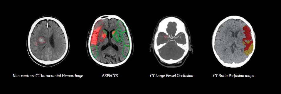 As part of the Automation Platform offering, innovative solution where created that helps optimize treatment outcomes for stroke patients when speed and accuracy are everything.
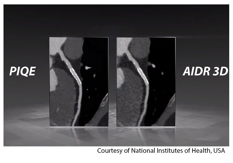
“We wanted to find a way to get the best of our two high-end CT scanners (the Aquilion ONE and Aquilion Precision) together, without huge cost, so as to provide the best value to our customers,” he said. “We think the solution is a particular technique called Super Resolution. Super Resolution is a type of algorithm that has been worked on for decades, but with the introduction of deep learning in recent years, it has advanced a great deal. It has, for example, been successfully applied in satellite imaging and in natural image processing on cell phones. So we wanted to bring this technology to CT and see what it could do. That's how we came up with the idea of a Super Resolution Deep Learning. And our resultant product is called Precise IQ Engine (PIQE).”
To train any neural network three ingredients are required: A training target that represents the ideal case, training input that represents the current system, and the neural network that can do the job.
Canon Medical has invested in all three components to optimize the algorithm and its performance. For the training target, Canon Medical has had the unique advantage of being able to access high resolution clinical data from the Aquilion Precision, which was introduced five years ago. This represents a wealth of high-quality clinical cases at a very good dose to present what the ground truth will look like. To create a training input the image is reconstructed at the highest resolution achievable. Denoising is implemented after, to allow us to preserve the best resolution possible. The neural network used is three-dimensional neural network.
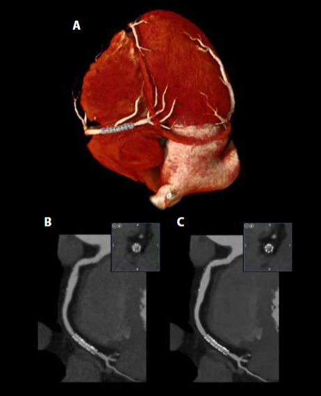
“In clinical cases, we have seen the benefits of PIQE, including improved visualization of stents, calcium, small vessels, aortic valves, and reduced blooming artifacts from calcium and stents,” remarked Dr. Yu. “All the doctors who have used PIQE agree that for the small structures that I have mentioned earlier, the conspicuity, the diagnostic confidence, has been greatly improved. I hope that this technology gets into the hands of more customers to enhance the care of patients.”
Optimized CT Workflow in Stroke with Deep Learning
The Diagnostic Image Analysis Group (DIAG) of Radboud University Medical Center in Nijmegen, the Netherlands, has been involved in the development and implementation of advanced imaging techniques that feature AI-solutions, in collaboration with Canon Medical for some time. One key focus is on the use of advanced neuroimaging tools to improve detection, diagnosis and treatment of stroke. In acute stroke, imaging needs to be performed and interpreted immediately, so that the right treatment for each patient can be determined and implemented fast. Recently the Medical Center has evaluated Canon Medical’s Automation Platform. Dr. Anton Meijer, Neuroradiologist provided an update on the applications around stroke, and workflow in particular.
“Time is of essence especially in stroke imaging so optimize CT workflow is crucial,” emphasized Dr. Meijer. “There are different points in the workflow which can be optimized with deep learning and AI.”
He explained that AI can also support the technician in selecting the correct scanning protocol to have image reconstruction either for optimized image quality but also to reduce radiation dose and can also support the physician in making the correct diagnosis, in stroke, with AI tools to instruct imaging for detection of hemorrhage or large vessel occlusions. He recognized that it can also support specialists in treatment decision-making.
“It is really important that we collaborate with the software engineers because the algorithm must be well trained and optimized,” he added. “ We must have adequate reference standards that should not only rely on the density of the abnormalities, especially in the skull base.”
“In stroke, it is important to have quick processing of perfusion CT. Deep learning really speeds up our workflow to make our diagnosis quick and fast and it is expected that also this CT perfusion acquisition will improve with deep learning processing, especially if we take clinical characteristics into account to have a more accurate prediction of outcome,” he said.
However, Dr. Meijer recognizes the challenges for the tools in clinical practice.
“The AI tools used in clinical practice must serve the clinicians and the radiologists,” he reiterated. “The ultimate goal is to improve the outcome of the patient, but for this we need real life outcome measures in order to have an idea what the added value of such tools are in clinical practice and often those are lacking in general, as is study of the effectiveness and value in clinical practice.”
“AI can facilitate the workflow which is really important for stroke imaging it can aid detection of relevant pathology and treatment decision-making, but it is really important that you know your AI tool that you manage expectations, and also that you evaluate your AI tool and collaborate with your with your vendors,” he concluded.
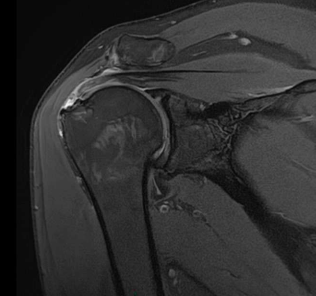
“With these high resolution scans we have very nice images and very good diagnosis,” he added. “When we perform an MRI-scan of the shoulder, we do this without contrast injection and we can see a lot of things in three planes very well. And then we decide if we require a direct arthrogram. I think we perform direct arthrograms in around 10-15% of cases, and this has improved a lot also with the introduction of Artificial Intelligence. We see much more detail on much better images. So, without contrast with high resolution images and Artificial Intelligence - it's very important for everything extra-articular, and we see the natural situation of the shoulder.”
Insight made possible
The ECR Satellite Symposium provided a broad spectrum of insights into how AI is successfully used in clinical practice, made possible by Altivity, Canon Medical’s bold new approach to AI innovation that uses smart technologies to make a whole new level of quality, insight, and value across the entire care pathway possible.
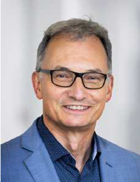
Prof. Mathias Prokop, MD, PhD
Radiologist/ Chairman of the Department of Radiology, Radboud University Medical Center University Medical Center Groningen Nijmegen, Groningen, the Netherlands.
Prof. Mathias Prokop, MD, PhD, is the department head of Imaging (Radiology, Nuclear Medicine and Anatomy) of Radboudumc in Nijmegen and the Department Head of Radiology at the University Medical Center Groningen in the Netherlands. He is the current president of the Dutch Society of Radiology (NVvR). Prof. Prokop is an expert in body imaging and multislice CT. Since 2004, he has focused on the early detection of disease, especially lung screening and cardiac imaging and thoracic applications of machine learning. His departments host one of the largest research teams in Europe. Besides main clinical groups that tackle lung, breast, prostate, and pancreatic cancer and metabolic disease and vascular malformations, the department's research covers ultrasound, CT, MRI, nuclear medicine, and AI, including computer-aided diagnosis and robot-assisted interventions.
Radiologist/ Chairman of the Department of Radiology, Radboud University Medical Center University Medical Center Groningen Nijmegen, Groningen, the Netherlands.
Prof. Mathias Prokop, MD, PhD, is the department head of Imaging (Radiology, Nuclear Medicine and Anatomy) of Radboudumc in Nijmegen and the Department Head of Radiology at the University Medical Center Groningen in the Netherlands. He is the current president of the Dutch Society of Radiology (NVvR). Prof. Prokop is an expert in body imaging and multislice CT. Since 2004, he has focused on the early detection of disease, especially lung screening and cardiac imaging and thoracic applications of machine learning. His departments host one of the largest research teams in Europe. Besides main clinical groups that tackle lung, breast, prostate, and pancreatic cancer and metabolic disease and vascular malformations, the department's research covers ultrasound, CT, MRI, nuclear medicine, and AI, including computer-aided diagnosis and robot-assisted interventions.
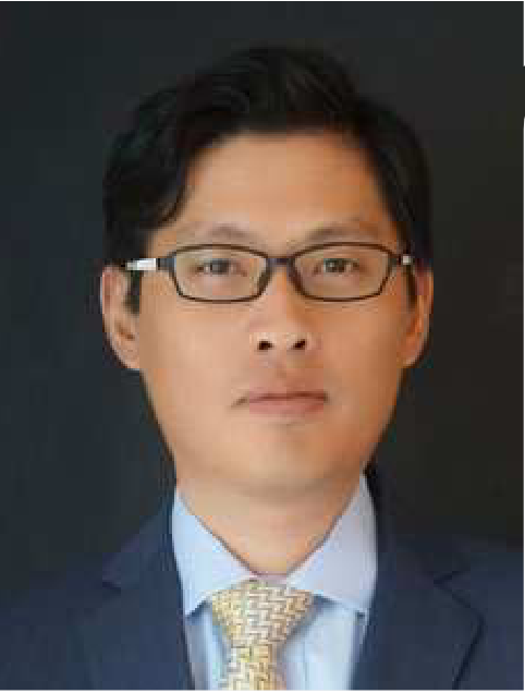
Dr. Zhou Yu, PhD
Director of CT R&D, Canon Medical Research USA, Inc., Chicago, USA.
Dr. Yu, Ph.D., is Director of CT R&D at Canon Medical Research USA, Inc. In this role, he leads the strategy and execution of CT research at CMRU. He manages a portfolio of advanced research and product development projects for Canon’s CT product lines. Zhou is a well-recognized expert on AI and deep learning image reconstruction and has more than 30 patents and 30+ peer-reviewed papers.
Director of CT R&D, Canon Medical Research USA, Inc., Chicago, USA.
Dr. Yu, Ph.D., is Director of CT R&D at Canon Medical Research USA, Inc. In this role, he leads the strategy and execution of CT research at CMRU. He manages a portfolio of advanced research and product development projects for Canon’s CT product lines. Zhou is a well-recognized expert on AI and deep learning image reconstruction and has more than 30 patents and 30+ peer-reviewed papers.
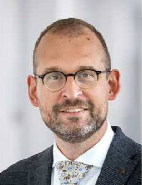
Dr. Anton Meijer, MD, PhD
Neuroradiologist, Radboud University Medical Center, Nijmegen, the Netherlands.
Dr. Anton Meijer is a Radiologist at the Department of Medical Imaging of Radboud University Medical Center, with a specialization in neuroradiology, and emergency radiology. He participates in clinical and research projects in neurovascular and neurodegenerative diseases. He is involved in the development and clinical implementation of advanced imaging techniques and AI solutions.
Neuroradiologist, Radboud University Medical Center, Nijmegen, the Netherlands.
Dr. Anton Meijer is a Radiologist at the Department of Medical Imaging of Radboud University Medical Center, with a specialization in neuroradiology, and emergency radiology. He participates in clinical and research projects in neurovascular and neurodegenerative diseases. He is involved in the development and clinical implementation of advanced imaging techniques and AI solutions.
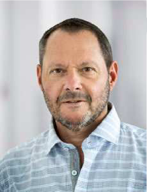
Dr. Jan Veryser, MD
Radiologist, Open MRI Zen, Sluis, the Netherlands.
Dr. Jan Veryser is the founder of Open MRI Zen, a private center in the Netherlands dedicated to musculoskeletal radiology. He is an active ESSR member and a member of the Imaging Guided Intervention Subcommittee. Dr. Veryser has a strong focus on musculoskeletal radiology and interventional ultrasound, especially in the field of nerve interventions. He regularly organizes workshops and lectures during international conventions (ECR, Arab Health, etc.) to improve the quality of ultrasound diagnosis and guided treatments.
Radiologist, Open MRI Zen, Sluis, the Netherlands.
Dr. Jan Veryser is the founder of Open MRI Zen, a private center in the Netherlands dedicated to musculoskeletal radiology. He is an active ESSR member and a member of the Imaging Guided Intervention Subcommittee. Dr. Veryser has a strong focus on musculoskeletal radiology and interventional ultrasound, especially in the field of nerve interventions. He regularly organizes workshops and lectures during international conventions (ECR, Arab Health, etc.) to improve the quality of ultrasound diagnosis and guided treatments.
Explore More

Prof. Mathias Prokop, MD, PhD
Radiologist/ Chairman of the Department of Radiology, Radboud University Medical Center University Medical Center Groningen Nijmegen, Groningen, the Netherlands.
Prof. Mathias Prokop, MD, PhD, is the department head of Imaging (Radiology, Nuclear Medicine and Anatomy) of Radboudumc in Nijmegen and the Department Head of Radiology at the University Medical Center Groningen in the Netherlands. He is the current president of the Dutch Society of Radiology (NVvR). Prof. Prokop is an expert in body imaging and multislice CT. Since 2004, he has focused on the early detection of disease, especially lung screening and cardiac imaging and thoracic applications of machine learning. His departments host one of the largest research teams in Europe. Besides main clinical groups that tackle lung, breast, prostate, and pancreatic cancer and metabolic disease and vascular malformations, the department's research covers ultrasound, CT, MRI, nuclear medicine, and AI, including computer-aided diagnosis and robot-assisted interventions.
Radiologist/ Chairman of the Department of Radiology, Radboud University Medical Center University Medical Center Groningen Nijmegen, Groningen, the Netherlands.
Prof. Mathias Prokop, MD, PhD, is the department head of Imaging (Radiology, Nuclear Medicine and Anatomy) of Radboudumc in Nijmegen and the Department Head of Radiology at the University Medical Center Groningen in the Netherlands. He is the current president of the Dutch Society of Radiology (NVvR). Prof. Prokop is an expert in body imaging and multislice CT. Since 2004, he has focused on the early detection of disease, especially lung screening and cardiac imaging and thoracic applications of machine learning. His departments host one of the largest research teams in Europe. Besides main clinical groups that tackle lung, breast, prostate, and pancreatic cancer and metabolic disease and vascular malformations, the department's research covers ultrasound, CT, MRI, nuclear medicine, and AI, including computer-aided diagnosis and robot-assisted interventions.
The content reflects information available at the time of publication and may differ from current information.
Social Media


