

Better Resolution = Better Patient Care
VISIONS spoke with Chief Physician Prof. Lukas Lusuardi and Senior Physician Dr. Martin Drerup, University Department of Urology and Andrology at Salzburg University Hospital, Austria.

“We frequently deal with pathologies of the outer genitals. Testicular carcinoma is the most common cancer in young men. The most important requirement sounds simple – to get an ultrasound image with the highest possible resolution,” says Dr. Drerup. The Xario 200G fully demonstrates its strengths in this respect. Not only is it compact and intuitive in its use, it also features exceptionally powerful imaging. The foundation of the Xario 200G is the High Density Beamformer, which enables technologies such as precision imaging and SMI as well as real-time compound imaging ApliPure+ and broadband harmonic imaging differential THI.
Image management, integrated onboard reporting and various network functions as well as up to 8 hours of battery life and a 2-second boot-up time, make the Xario 200G a high-performance ‘team member’ in the urology department and a perfect partner for all-around tasks. Together with the Aplio i700, clinical requirements can be optimally fulfilled.
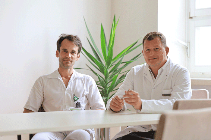
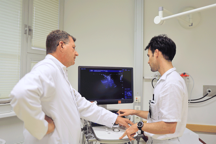
Fusion biopsy for prostate cancer is a frequent area of application for ultrasound. “The objective is to find the best treatment for each individual patient,” says Prof. Lusuardi. And this works best with an MRI-guided fusion biopsy, during which tissue samples which were previously remarkable on the MRI scan are removed in a targeted manner using ultrasound imaging.
To improve the accuracy of the exam, the MRI and ultrasound images are superimposed on each other. The MRI scan serves as ‘visual support’ to permit improved navigation within the higher-resolution ultrasound visualisation. Due to the accuracy, not only can we detect a higher number of clinically relevant tumours, we can also reduce the amount of biopsies. //

Senior Physician Dr. Martin Drerup,
University Department of Urology and Andrology at Salzburg University Hospital,
Austria.
University Department of Urology and Andrology at Salzburg University Hospital,
Austria.
Chief Physician Prof. Lukas Lusuardi,
University Department of Urology and Andrology at Salzburg University Hospital,
Austria.
University Department of Urology and Andrology at Salzburg University Hospital,
Austria.
Where do you see the current importance of ultrasound diagnostics within the context of new technical developments of other imaging procedures?
Prof. Lusuardi: Thanks to its fast diagnostic capability and availability, the importance of ultrasound in urology is constantly increasing. Ultrasound is quite simply a workhorse – for example, in the daily routine at the ward, not a lot will change with respect to follow-ups in the post-operative setting.
Which trends and developments do you see in the area of ultrasound diagnostics for urology?
Prof. Lusuardi: That’s quite clearly fusion biopsy. For outer genital pathologies, ultrasound will remain the method of choice despite improved cross-sectional imaging. The same applies to kidney stone removal, in my view. In the core urology areas, ultrasound won’t soon be replaced by cross-sectional imaging.
What are your requirements for ultrasound equipment in general?
Dr. Drerup: We’ve compared multiple devices, and intuitive operation is top priority. And another requirement was recently added: The equipment components must be easy to clean and/or disinfect.Which requests would you have for the application team or what type of support do you expect for your clinical routine?
Dr. Drerup: The fundamental standard requirements – that is, how the device is operated – need to be demonstrated to every employee. And then there are the special procedures, such as fusion biopsies, where we would like an application expert to be an expert in this field as well. The Austrian team additionally sent us a specialist from their European headquarters for this. That was very helpful for all of us, and we were able to have high-level discussions about the application.What are the requirements for future ultrasound diagnostics and hence for your colleagues’ education and training?
Prof. Lusuardi: There is a big difference between theory and practice. We would hope that everyone receives the same education and training. But in reality it’s not always possible during the residency rotation period for everyone to be trained in all urology aspects equally well and for the same amount of time.The content reflects information available at the time of publication and may differ from current information.
Social Media


