


Latest news from our
VISIONS Magazine
Offering a Wealth of Unexplored Possibilities
January 24, 2022
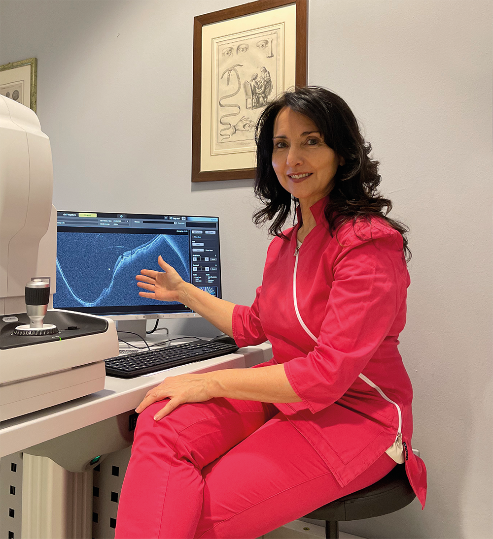
VISIONS spoke with Dr. Barbara Parolini (Head of the Vitreo Retinal Unit at the Eyecare Clinic, Brescia, Italy) about Canon's Xephilio OCT-S1 and OCT-A1 Eye Care systems.
Dr. Barbara Parolini is Head of the Vitreo Retinal Unit at the Eyecare Clinic, a private institution in Brescia, Northern Italy. In an interview with Visions, she explained how the Xephilio OCT-S1 and OCT-A1 enable her to better understand pathology, confirm suspected disease and follow up patients even remotely.
Biography
Dr. Barbara Parolini was born on April 17th, 1968 in Italy. She underwent her postodoctoral fellowship in 1994-1995 at UCSF (San Francisco, USA) and in 1997-1998 at UMDNJ (Newark, USA).
She is Head of Vitreo Retinal Unit at Eyecare Clinic, in Brescia, Italy. She has treated 10,000 cases of complex cases of vitreoretinal pathologies in both adult and paediatric patients; submacular surgery; high myopia; cataract; glaucoma and refractive surgery.
She has published 60 peer-reviewed papers and four book chapters.
She’s a tutor of the Bremen Vitreo Retinal School and the Thessaloniki Vitreo Retinal School.
She’s regularly invited as speaker and moderator, and to perform live surgery all around the world.
She’s a reviewer for multiple peer-reviewed journals and Vice President of the European Vitreoretinal Society (EVRS).
www.retinitaly.net is an educational website for patients and doctors with videos
Dr. Barbara Parolini was born on April 17th, 1968 in Italy. She underwent her postodoctoral fellowship in 1994-1995 at UCSF (San Francisco, USA) and in 1997-1998 at UMDNJ (Newark, USA).
She is Head of Vitreo Retinal Unit at Eyecare Clinic, in Brescia, Italy. She has treated 10,000 cases of complex cases of vitreoretinal pathologies in both adult and paediatric patients; submacular surgery; high myopia; cataract; glaucoma and refractive surgery.
She has published 60 peer-reviewed papers and four book chapters.
She’s a tutor of the Bremen Vitreo Retinal School and the Thessaloniki Vitreo Retinal School.
She’s regularly invited as speaker and moderator, and to perform live surgery all around the world.
She’s a reviewer for multiple peer-reviewed journals and Vice President of the European Vitreoretinal Society (EVRS).
www.retinitaly.net is an educational website for patients and doctors with videos
Welcome to Brescia, ‘the Lioness of Italy’, home of Italian caviar. This beautiful town, more discreet than its neighbor Verona, attracts sightseers looking for something new. Some travel a long way for the opportunity to materialize a vision: improve their eyesight.
Many of these visitors are patients of the Eyecare Clinic, where Dr. Parolini works with the finest Canon Medical equipment.
“I see about 30 patients per day. Most patients have been referred from all over the country and abroad for retinal pathologies, and peculiar cataract and refractive procedures,” she said.
Dr. Parolini and her team provide full eye check, central nerve and macula OCT, ultrawide OCT, angio OCT and anterior segment OCT as well as central and wide retinography, autofluorescence, and, when needed, microperimetry, corneal map, biometry and endothelial cell count.
To support their procedures with optimal imaging technology, Dr. Parolini and her three orthoptics colleagues use the Xephilio OCT-A1 and OCT-S1. The latter was installed in July 2020 and has become essential to daily practice.
“The challenge was to fit both machines into the routine workflow. That’s why I have three technicians working with me now,” she said.
“I want as much imaging as I can get for each patient. Imaging is essential for collecting precise information to reach a correct diagnosis during and after consultation. I study the most complicated cases even after the patient is gone and compare my images with published data. I also use images for educational purposes.”
“Working with the Xephilio OCT-S1 and OCT-A1 offers plenty of benefits, starting with a better understanding of the pathologies.”
Dr. Barbara Parolini.
Improving daily routine and bringing new opportunities
“Working with the Xephilio OCT-S1 and OCT-A1 offers plenty of benefits, starting with a better understanding of the pathologies,” she said. The equipment also helps confirm suspected pathology and follow up patients, both in the conventional and telemedicine setting.
Having to diagnose patients remotely is a frequent scenario, especially in the case of an emergency. “My orthoptists are such experts now that they scan the fundus with color wide field retino-graphy and wide field OCT and send me the images in real time. So, I can decide which management to offer even when I’m not at the clinic. This is a real optimization of my work and a great plus for the clinic and the patients.”
The technology enables to see further than conventional techniques. Dr. Parolini recently diagnosed peripheral schisis and retinal detachments that were invisible using a contact lens, with the patient sitting at the slit lamp, or wide field retinography. “I was astonished myself. Using wide-field OCT, I realize how much more we can see.”
Another key advantage brought by the Xephilio is the study of the retina in non-cooperative patients, such as children and patients with high photopsia. “The light of the OCT is much easier to endure for patients.
And with the imaging of the OCT-S1, I have enough information to check almost every pathology on any patient. I can collect great images full of details even in miosis, which is great for patients with shallow anterior chamber or suspect allergies and pregnant women.”
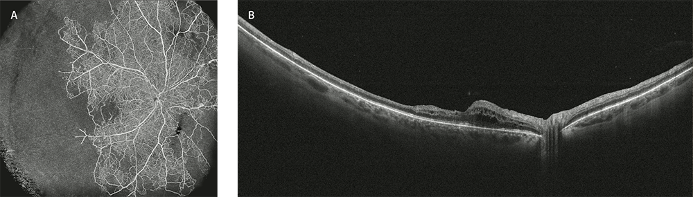
Central retinal vein occlusion
A) AngioOCT of the superficial capillary plexus with Canon S1. Absence of blood flow in the temporal inferior and nasal quadrants.
B) OCT Bscan. Thinning of the temporal retina and macular edema of the nasal macula.
Canon’s high-end technology brings many improvements in daily routine, allowing higher resolution of images, wider scans, images superimposed on the retinography, faster angiography and enface OCT of the retina layer.
“The equipment provides new insights of the peripheral area of retina, vitreous and choroid,” she said.
As Vice President of the European Vitreoretinal Society (EVRS), a pro-fessional independent initiative, Dr. Parolini is keen on sharing knowledge and detecting new trends in medical retina surgery and imaging.
“The equipment provides new insights of the peripheral area of retina, vitreous and choroid,” she said.
As Vice President of the European Vitreoretinal Society (EVRS), a pro-fessional independent initiative, Dr. Parolini is keen on sharing knowledge and detecting new trends in medical retina surgery and imaging.
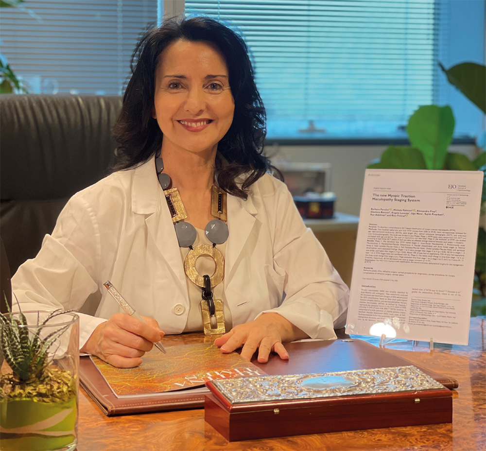
“Using wide-field OCT, I realize how much more we can see.”
Dr. Barbara Parolini
She expects the OCT-S1 and OCT-A1 to push wide field angiography so that it can replace fluorescent angiography whenever possible. Angiography of the choroid will also advance in the future thanks to the combined efforts of clinicians, researchers and the industry.
“The Xephilio will change patient management and enable to image every patient in the periphery of the retina with more precise diagnosis,” she said.
Working with Canon Medical Systems has proved a wonderful experience. “It’s been an extremely smooth, fast, and enthusiastic collaboration. I appreciated the approach by the company and would love to undertake multicenter studies through the Canon network.”
A passionate educator and tutor for the European Vitreo Retinal schools in Bremen, Germany and Thessaloniki, Greece for many years, Dr. Parolini sees cooperation as an essential part of her work. “I always love comparing my way with the others’. I love to learn endlessly.” //
“The equipment provides new insights of the peripheral area of the retina, vitreous and choroid.”
Dr. Barbara Parolini
Other blogs that may interest you
Social Media


