


Latest news from our
VISIONS Magazine
Leveraging Additional Layers of Intelligence
October 10th, 2023
Research-led advances in healthcare practices sometimes requires the acquisition of new technology. One recent example is the adoption of new guidelines on stroke assessment protocols in the Netherlands following integration of results from the MR CLEAN study1 – a trial of intra-arterial treatment for acute ischemic stroke, and the MR CLEAN-LATE study2. These studies provided new insights in the treatment of ischemic stroke made possible through the use of CT brain perfusion after brain infarctions. This imaging technique was found to give a good indication of brain tissue status before deciding the best options for neuro intervention or thrombolysis.
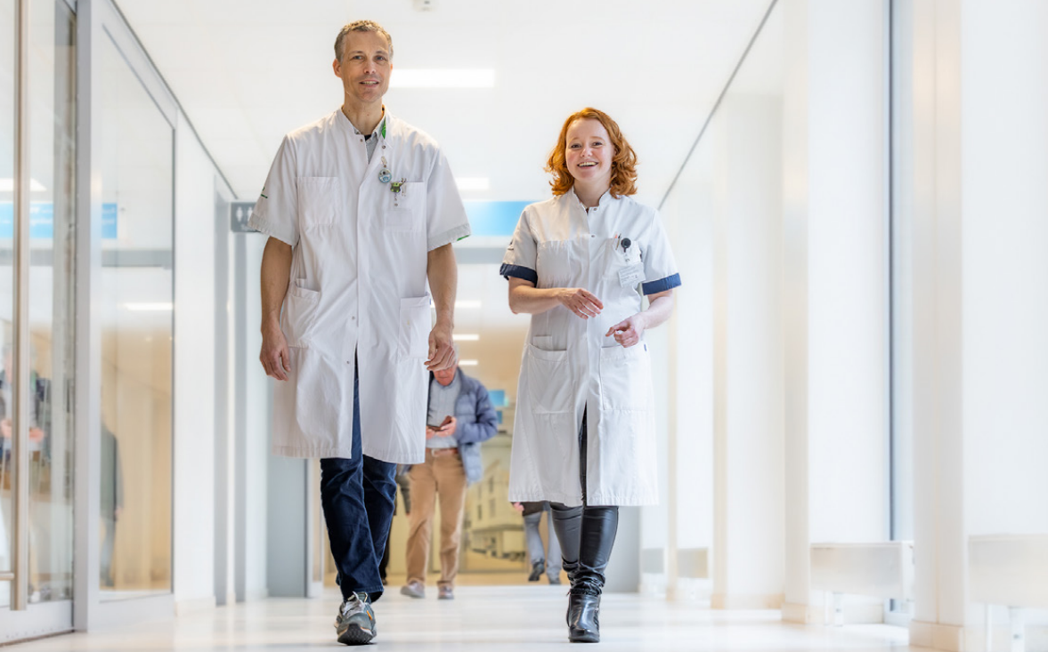
With the introduction of new protocols across the Netherlands in 2021, the Groene Hart Ziekenhuis (GHZ) in Gouda, the Netherlands, was able to fulfill the new guidelines thanks to Canon Medical technology. Stijn Bollen, Radiologist, and Melissa van den Bos, Medical Physicist Expert at the GHZ, explain how the systems and software assist their work.
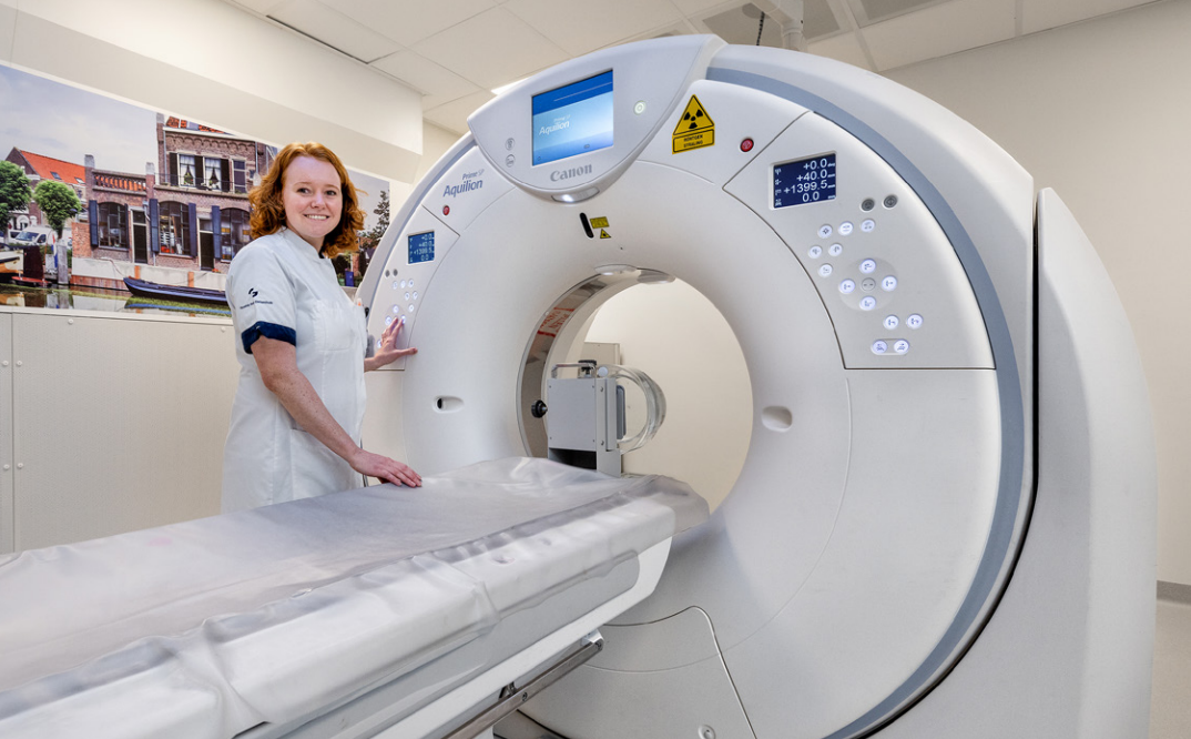
GHZ is a general hospital that offers state-of-the-art medical care from four different locations, from which Gouda is the main location (Gouda, Bodegraven, Schoonhoven, and Zuidplas) all of which lie within the “Groene Hart” (Green Heart) area of the Netherlands. It has two Canon Medical Aquilion Prime SP CT scanners with Vitrea Advanced Visualization and Automation Platform.
“At the end of 2021, we implemented the brain perfusion CT protocol in addition to CT angiography for detecting acute infarctions and calculating the percentage of penumbra and core, to follow the new updated Dutch guidelines,” said Stijn. “With implementation of this new protocol, we also started to use the Automation Platform from Canon Medical to confirm the diagnosis of ischemic stroke and to estimate tissue viability.”
“During the day and night, we scan patients with symptoms of brain infarction, therefore, we make use of Canon Medical’s Automation Platform,” added Stijn.
 * when Automation Platform is mentioned in the paragraph above the disclaimer.
New perspectives
GHZ opted for the Automation Platform because it helps their radiologists obtain brain perfusion maps for direct assessment, and enable fast indication of Large Vessel Occlusion (LVO) and detection of Intracranial Hemorrhage (ICH).
“After performing a CT brain perfusion scan, the CT slices are sent to the Automation Platform for analysis. The analyzed brain perfusion maps, including indication of large vessel occlusion or intracranial hemorrhage, are saved and shown in PACS, consistent with normal workflow for CT imaging,” explained Melissa.
GHZ opted for the Automation Platform because it helps their radiologists obtain brain perfusion maps for direct assessment, and enable fast indication of Large Vessel Occlusion (LVO) and detection of Intracranial Hemorrhage (ICH).
“After performing a CT brain perfusion scan, the CT slices are sent to the Automation Platform for analysis. The analyzed brain perfusion maps, including indication of large vessel occlusion or intracranial hemorrhage, are saved and shown in PACS, consistent with normal workflow for CT imaging,” explained Melissa.

“The greatest impact of the Automation Platform for the team is seen during night-time, due to the quick availability of the images.”
Stijn Bollen, Radiologist, the Groene Hart Ziekenhuis (GHZ), Gouda, the Netherlands.
“They provide observer-independent analysis/volume determination,” continued Melissa. “The analysis of brain perfusion maps created by Automation Platform correspond with manually analyzed brain perfusion images of patients.”
GHZ have undertaken their own studies to assess the results obtained by the Automation Platform. They compared the penumbra and core volumes analyzed by the Automation Platform with manual analysis using the Vitrea Advanced Visualization workstation and found no large differences.
The neuroradiologists at GHZ also use the analyzed brain perfusion maps from the Automation Platform.
Benefits of ICH and LVO algorithms
Previously, radiologists could only rely on analysis of the unenhanced CT brain scan for evidence of an infarction, mass, or hemorrhage, and CT Angiography of the brain from which to find vessel occlusions or other abnormalities.
The introduction of Canon Medical’s Automation Platform provides extra tools to exclude hemorrhage and large vessel occlusion through its ICH and LVO algorithms.
“Within the context of ‘time is brain’ in diagnosing patients with symptoms of brain infarction, Automation Platform has definitely improved the speed and quality of diagnosis in acute stroke,” said Stijn. “The main diagnostic advantage is the LVO/ ICH indication on the native and CTA images.”
“It gives an additional value in the level of confidence for the radiologist, “continued Stijn. “When there is a perfusion defect, with no acute occlusion or spasm, the radiologist is more focused on finding alternative causes, such as spasm or smaller peripheral occlusions.”

“Vitrea and Automation Platform are stable and reliable software systems within the hospital.”
Melissa van den Bos, Medical Physicist Expert, the Groene Hart Ziekenhuis (GHZ), Gouda, the Netherlands.
Perfusion maps
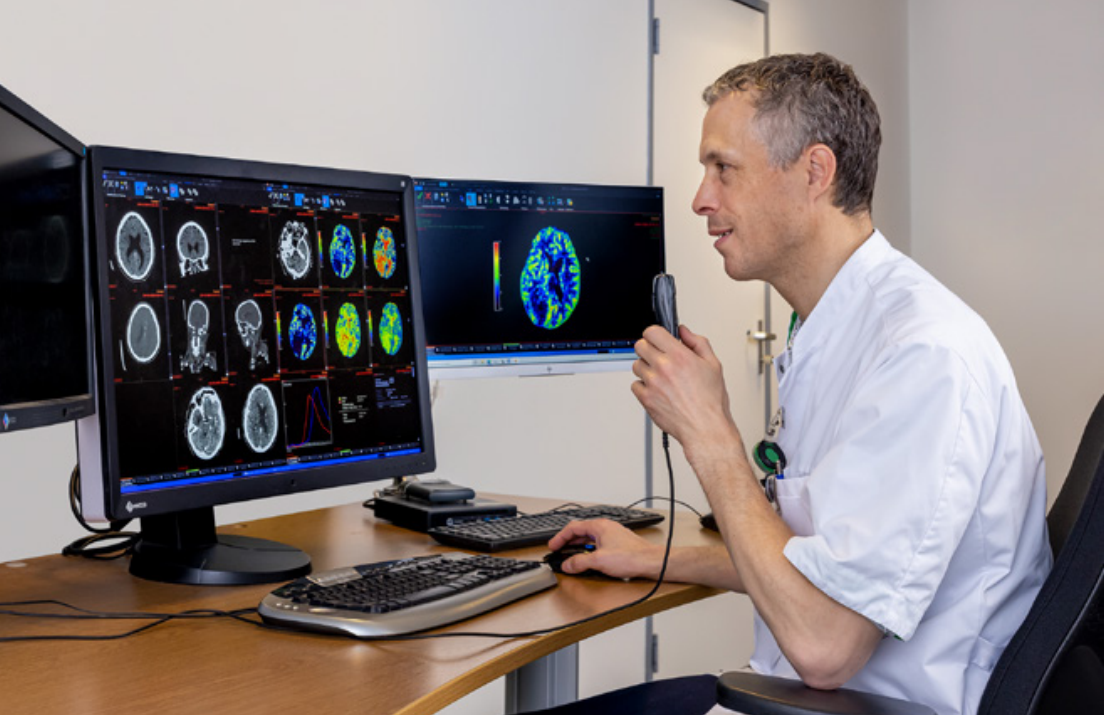
Using the systems, the radiologists are able to obtain CT Perfusion (CTP) maps that show CBV (Cerebral Blood Volume), CBF (Cerebral Blood Flow), MTT (Mean Transit Time), and Tmax (Time to maximum).
“The CTP maps gives an indication of whether an endovascular therapy, such as intra-arterial thrombolysis in a later time window would still be useful,” explained Stijn. “The maps are accurate. The workflow improves because there is no need to open the Vitrea Advanced Visualization to obtain the perfusion maps.”
Full training
When the Vitrea Advanced Visualization and Automation Platform were initially installed, the radiologists at GHZ received training on the brain perfusion maps and calculating volumes. Training on the Automation Platform was provided to the hospital’s radiologists and some CT technicians when it was operational. And the radiologists received additional training in learning how to access and interpret the extra images, which include Large Vessel Occlusion (LVO) and Intracerebral Hemorrhage (ICH) indication. The CT technicians also received training for the scan protocol for perfusion scans.
“We have a good relationship with the Canon Medical Team,” remarked Melissa. “The technicians gave good support when the Aquilion Prime SP CT, Vitrea Advanced Visualization and Automation Platform systems were introduced, and they continue to provide great support, for example, when we have questions. We were able to transfer their know-how in the form of excellent support.”
Making new protocols easy
Canon Medical’s Vitrea Advanced Visualization and Automation Platform systems have not only enabled GHZ to integrate the new Dutch guidelines on assessment of stroke patients into clinical practice, but they provide ease of use that has improved workflow and diagnostic confidence.
Using the systems, the radiologists are able to obtain CT Perfusion (CTP) maps that show CBV (Cerebral Blood Volume), CBF (Cerebral Blood Flow), MTT (Mean Transit Time), and Tmax (Time to maximum).
“The CTP maps gives an indication of whether an endovascular therapy, such as intra-arterial thrombolysis in a later time window would still be useful,” explained Stijn. “The maps are accurate. The workflow improves because there is no need to open the Vitrea Advanced Visualization to obtain the perfusion maps.”
Full training
When the Vitrea Advanced Visualization and Automation Platform were initially installed, the radiologists at GHZ received training on the brain perfusion maps and calculating volumes. Training on the Automation Platform was provided to the hospital’s radiologists and some CT technicians when it was operational. And the radiologists received additional training in learning how to access and interpret the extra images, which include Large Vessel Occlusion (LVO) and Intracerebral Hemorrhage (ICH) indication. The CT technicians also received training for the scan protocol for perfusion scans.
“We have a good relationship with the Canon Medical Team,” remarked Melissa. “The technicians gave good support when the Aquilion Prime SP CT, Vitrea Advanced Visualization and Automation Platform systems were introduced, and they continue to provide great support, for example, when we have questions. We were able to transfer their know-how in the form of excellent support.”
Making new protocols easy
Canon Medical’s Vitrea Advanced Visualization and Automation Platform systems have not only enabled GHZ to integrate the new Dutch guidelines on assessment of stroke patients into clinical practice, but they provide ease of use that has improved workflow and diagnostic confidence.

You can also download this article as PDF or the full edition of the respective VISIONS
Other blogs that may interest you
Social Media


