


Latest news from our
VISIONS Magazine
CT Examination of Three Symptomatic Wild Seals
February 28th, 2024
Seals are wild animals and are, therefore, rarely brought to vets when they are sick. One exception from this is seen at seal sanctuaries, which administer medical care to sick or young stranded seals that have been rescued and admitted. An example of this is the nonprofit organization, A-Seal, in Stellendam, the Netherlands. This organization rescues and rehabilitates seals that have been stranded along the nearby Dutch coastline. These seals are often pups that have been separated from their mothers, but they can also be young or adult seals that are sick or injured. When treatment and rehabilitation are successful and complete, the seals are released back into the wild.
Veterinarians face various challenges when they examine wild seals because they are often not cooperative. Other practical problems are encountered due to seals’ stocky build and thick layer of blubber, which can make manual examination difficult, especially of the spine, limbs or joints. In cases of a suspected ear problem, otoscopy can also be very difficult to perform, as seals have a very narrow external auditory canal.
To overcome some of these problems, radiographic examinations are often carried out in rehabilitation centers, mainly of bony structures and the lungs. However, in some patients a CT scan can be a quick and non-invasive way to obtain additional and more detailed information.
The following series of clinical cases illustrates the importance of performing CT scanning in wildlife. The cases demonstrate that CT is suitable for assessing anatomical structures that are not clearly visible on plain radiography.
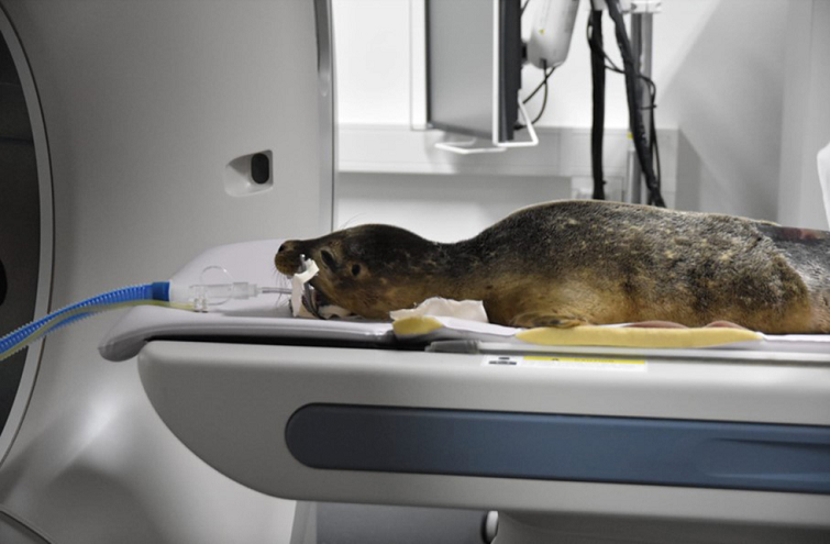
Three young seals, recently rescued on the beach, were examined by the responsible veterinarian, Dr. Machteld Geut. The clinical findings of the three cases were then discussed with veterinarians, Dr. Gert ter Haar and Dr. Susanne A.E.B. Boroffka. This multidisciplinary consultation concluded that CT examination is necessary for optimal imaging and evaluation of the submitted seals.
 None
None
Anesthesia
Specific problems can arise during anesthesia in pinnipeds due to the anatomical and physiological adjustments they have for long-term diving. The so-called 'dive response' causes apnea and deep bradycardia. The same apnea response is often observed in combination with treatment or during anesthesia. This can lead to hypoxemia and in severe cases to cardiac arrest1. A special anesthesia protocol developed by Geraldine Lacave, marine mammal veterinarian, was used.
All seals were first anesthetized with an intramuscular injection (butorphanol).
Specific problems can arise during anesthesia in pinnipeds due to the anatomical and physiological adjustments they have for long-term diving. The so-called 'dive response' causes apnea and deep bradycardia. The same apnea response is often observed in combination with treatment or during anesthesia. This can lead to hypoxemia and in severe cases to cardiac arrest1. A special anesthesia protocol developed by Geraldine Lacave, marine mammal veterinarian, was used.
All seals were first anesthetized with an intramuscular injection (butorphanol).
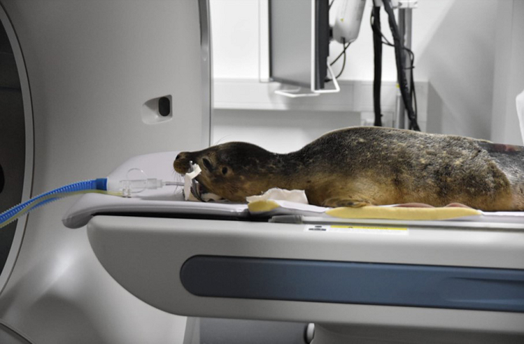
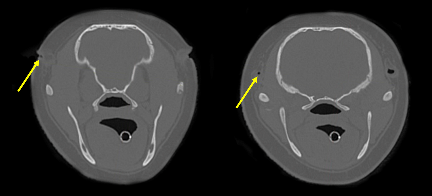 Figure 1a and 1b show the patient's ear canal (yellow arrows) in bone filter reconstructions. The transverse images show the narrow ear canal (a) and the obvious narrowing of the left external ear canal (b).
After they were sufficiently anesthetized, the induction medication (propofol) was administered via the extradural epidural vertebral vein (vertebral sinus) in the lumbar back, which is well-developed in all pinnipeds.
After induction, the seals were intubated and connected to a loop ventilation system with mechanical ventilation. Anesthesia was maintained with isoflurane in oxygen. The anesthetized seals were then taken to the CT room and placed in sternal position on the CT table. Pre- and post-contrast CT examinations were performed over the entire body.
Seal 01: Vincent
Vincent presented with chronic purulent discharge from his left ear. Therefore, it was particularly important to evaluate the external auditory canal, middle and inner ear. Seals do not have an external auricle; they have a small opening that closes underwater.
After induction, the seals were intubated and connected to a loop ventilation system with mechanical ventilation. Anesthesia was maintained with isoflurane in oxygen. The anesthetized seals were then taken to the CT room and placed in sternal position on the CT table. Pre- and post-contrast CT examinations were performed over the entire body.
Seal 01: Vincent
Vincent presented with chronic purulent discharge from his left ear. Therefore, it was particularly important to evaluate the external auditory canal, middle and inner ear. Seals do not have an external auricle; they have a small opening that closes underwater.
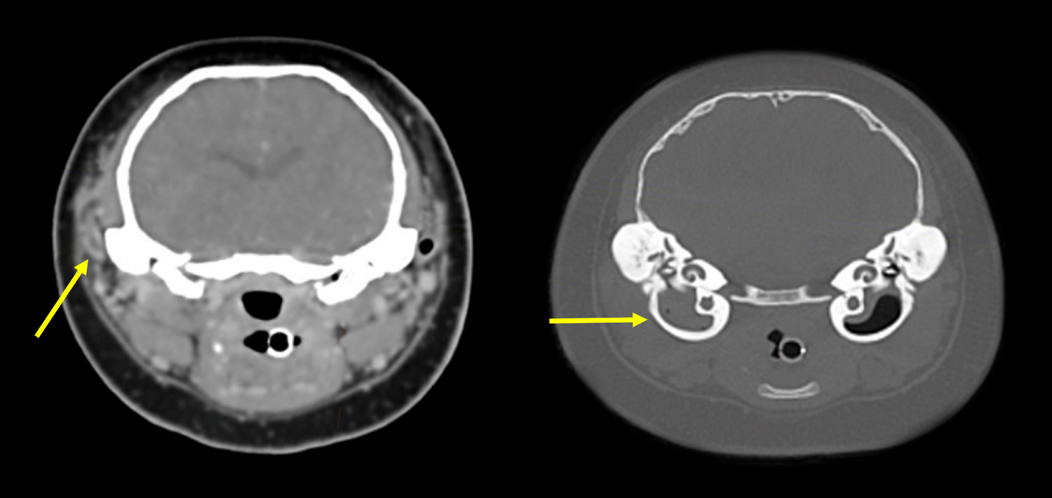 Figure 1c and 1d. Image 1c shows the blocked ear canal in the soft tissue filter reconstruction and image 1d shows the fluid-filled bulla in the bone filter reconstruction.
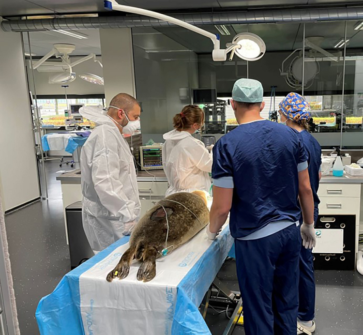
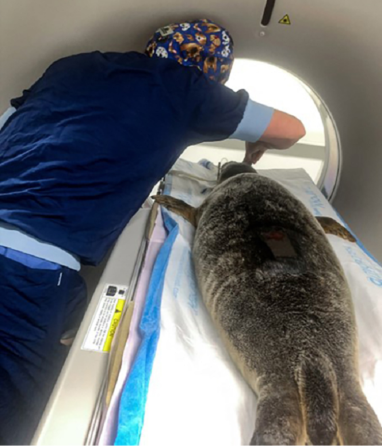
The CT examination showed a clear complete closure of the left external auditory canal due to thickening of soft tissue (Figure 1). Moreover, the left tympanic bulla was filled with soft tissue. The right bulla was filled with air, as normal.
After administration of contrast fluid, a contrast enhancement of the slightly thickened wall of the left external auditory canal was observed, but the contents of the cavity did not suggest an accumulation of fluid, cerumen or mucus. The contents of the left bulla showed no contrast enhancement.
The narrowed external auditory canal and irregular wall thickening corresponded with an external ear infection. Vincent underwent medical treatment for the external ear infection several times without success. After a multidisciplinary consultation, it was decided that he should undergo surgery for full recovery. After surgery and recovery, Vincent was returned to A-Seal, where he was kept out of the water for several days before being released into a pool of salt water.
Seal 02: Cascada
Cascada presented with a lop-sided head positioning indicating an inner ear problem, but without any discharge from the ears. A CT examination was performed to rule out otitis media/otitis interna. Transverse and dorsal CT images show normal air attenuation in both tympanic bullae (Figure 2). Based on the CT scan, a middle ear infection was ruled out.
After administration of contrast fluid, a contrast enhancement of the slightly thickened wall of the left external auditory canal was observed, but the contents of the cavity did not suggest an accumulation of fluid, cerumen or mucus. The contents of the left bulla showed no contrast enhancement.
The narrowed external auditory canal and irregular wall thickening corresponded with an external ear infection. Vincent underwent medical treatment for the external ear infection several times without success. After a multidisciplinary consultation, it was decided that he should undergo surgery for full recovery. After surgery and recovery, Vincent was returned to A-Seal, where he was kept out of the water for several days before being released into a pool of salt water.
Seal 02: Cascada
Cascada presented with a lop-sided head positioning indicating an inner ear problem, but without any discharge from the ears. A CT examination was performed to rule out otitis media/otitis interna. Transverse and dorsal CT images show normal air attenuation in both tympanic bullae (Figure 2). Based on the CT scan, a middle ear infection was ruled out.
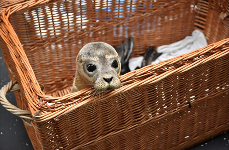
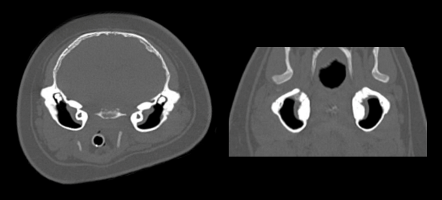 Figure 2: No abnormalities were observed on both bone filter reconstructions.
In the diagnosis of health problems in wildlife, there is usually no case history to work from. Examination and treatment of wild animals is not as easy as in domestic animals that are used to being handled. In addition, the conditions that affect wildlife can be very specific, even species’ specific, due to their physiology and habitat. In the case of wildlife rescue, resources such as veterinary care are often very limited, which places further emphasis on getting diagnoses and treatment right first time. Undoubtedly, CT has an important role in optimizing wildlife healthcare.
Drs. Bas Wetzels, Resident ECVDI
Drs. Machteld Geut, Veterinarian specialized in marine mammals
Dr. Susanne A.E.B. Boroffka, PhD, DECVDI
Dr. Gert ter Haar, PhD, DECVS
Drs. Bas Wetzels, Resident ECVDI
Drs. Machteld Geut, Veterinarian specialized in marine mammals
Dr. Susanne A.E.B. Boroffka, PhD, DECVDI
Dr. Gert ter Haar, PhD, DECVS
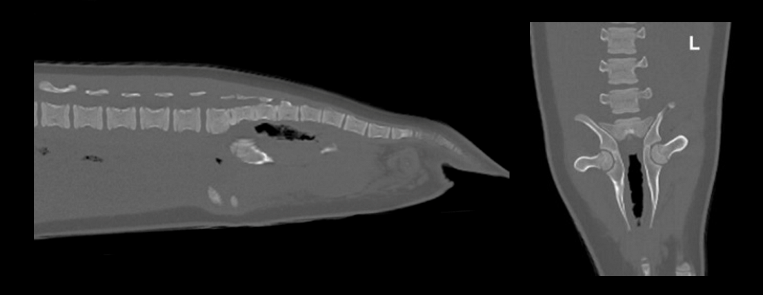 Figure 3: No abnormalities were seen in either view of the bone filter reconstructions of the spine and pelvis in Elvis.
Other blogs that may interest you
References
1 Huuskonen, V., Hughes, L. & Bennett, R. Anaesthesia of three young grey seals (Halichoerus grypus) for fracture repair. Irish Vet J 64, 3–3 (2011).
Social Media


