

ADVANCES IN OPHTHALMOLOGY - Wide Field OCTA May Revolutionize Retinal Vascular Disease Care
The use of eye examinations as reliable predictive tools for a range of conditions in healthcare is expanding. Several different image modalities give information about structural and functional vascular information in the eye, and eye examinations are fast and convenient. Examination with Optical Coherence Tomography Angiography (OCTA) is non-invasive, does not require fluorescein injection or pupil dilation, and takes just seconds.
In the last couple of years, wide field OCTA technology has come to the forefront in its potential to transform the assessment of retinal vasculature. Manchester Royal Eye Hospital in the United Kingdom (UK) was the first in the country to utilize Canon Medical System’s new, ultra-wide field OCTA system – the Xephilio OCT-S1 – in a patient-facing setting. Professor Tariq Aslam, Professor of Ophthalmology and Consultant Ophthalmologist at the hospital, explained to VISIONS how the system holds significant advantages for imaging and managing the impact of diabetes on the eye.
A growing problem
More than 830 million people across the world have diabetes. This number has consistently grown for decades. Early screening for diabetes-related changes is essential for effectively managing the condition and reducing the risk of serious complications, including those that affect retinal health.
“With the rising prevalence of diabetes, effective and efficient management of diabetic retinopathy remains a logistical and clinical challenge,” explained Prof. Aslam. “It requires multiple visits for monitoring and treatment with multiple potential modes of imaging that can include invasive and intravenous dyes. There are multiple potential avenues for treatment, such as laser treatments, anti-VEGF and steroids, as well as surgical interventions and systemic control.”

“Wide field OCTA and the Xephilio OCT-S1 have definite potential to revolutionize the care of patients with retinal vascular disease.”
Prof. Tariq Aslam.
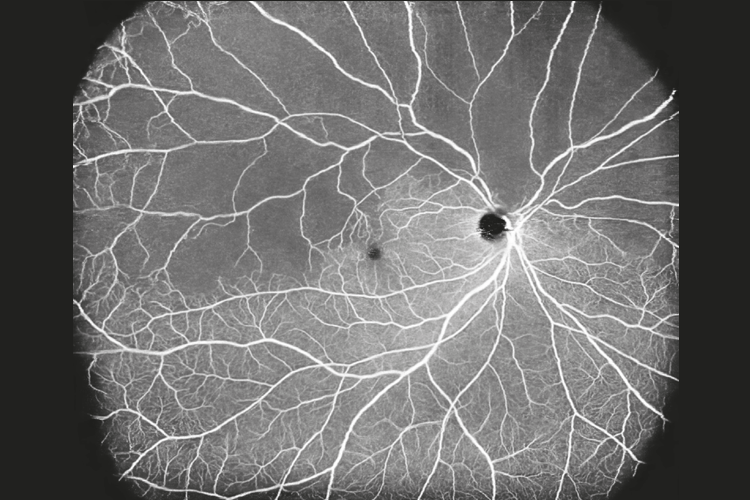
“The main advantages of wide field OCTA over traditional imaging methods are convenience and accessibility,” he remarked. “The scans can easily be done at every visit, and available or comparative assessments before each consultation. This is not possible with fluorescein angiography.”
“The information it provides does not completely replace the current gold standard of fluorescein angiography, but in many key areas required for decision-making for fundamental questions, such as the risk of neovascularization, there is a rising body of evidence that wide field OCTA provides a pragmatic alternative for a majority of cases,” he explained. “It may, furthermore, provide information that other traditional systems cannot, such as information on different depths of networks and vessel intensity, as well as a great ease of application of image processing and analysis for machine learning and expert algorithms.”
Working with the Xephilio OCT-S1 has enabled Prof. Aslam to detect neovascularization more clearly and supported monitoring of new vessels, which has allowed for more confident, sensitive and specific use of treatments.
“In the future, studies with the system may be able to allow for prediction of the likelihood of progression to later stages from early scans,” he added. “We are currently exploring the potential of the Xephilio OCT-S1, as well as adaptive optics, in the early detection of Alzheimer’s Disease. However, there are also a large number of systemic diseases that might have potential for input from the data these scans provide.”
“The information it provides does not completely replace the current gold standard of fluorescein angiography, but in many key areas required for decision-making for fundamental questions, such as the risk of neovascularization, there is a rising body of evidence that wide field OCTA provides a pragmatic alternative for a majority of cases,” he explained. “It may, furthermore, provide information that other traditional systems cannot, such as information on different depths of networks and vessel intensity, as well as a great ease of application of image processing and analysis for machine learning and expert algorithms.”
Working with the Xephilio OCT-S1 has enabled Prof. Aslam to detect neovascularization more clearly and supported monitoring of new vessels, which has allowed for more confident, sensitive and specific use of treatments.
“In the future, studies with the system may be able to allow for prediction of the likelihood of progression to later stages from early scans,” he added. “We are currently exploring the potential of the Xephilio OCT-S1, as well as adaptive optics, in the early detection of Alzheimer’s Disease. However, there are also a large number of systemic diseases that might have potential for input from the data these scans provide.”
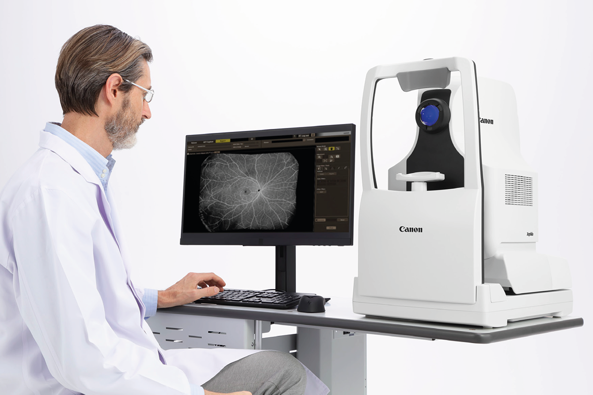
AI-based image processing provides enhanced clarity
Prof. Aslam has found the Xephilio OCT-S1’s sophisticated, AI supported, image post-processing with 3D projection artefact removal ensures excellent image quality.
“OCTA can be harder to interpret than standard OCT due to the complexity of the images and additional dimensions and artifacts introduced, requiring a dynamic assessment of multimodal OCTA outputs, including enface scans of different layers, and B scans with flow signals,” he said., “However, the user interface of the Xephilio OCT-S1 is convenient and practical to allow for this and the artificial intelligence-enhanced images also improve clarity.
Smooth adoption
Canon Medical Systems and SenseMedical supported Prof. Aslam and his team throughout the adoption and implementation of the Xephilio OCT-S1.
“There are specific needs for NHS procurement, data storage, screen resolutions, computer terminal capacity and so on, in the UK, that Canon has helped us ensure were adequate to allow us to acquire, and then fully utilize the imaging,” he explained.
Potential to revolutionize care for retinal vascular disease
Through research and use in clinical practice for more than one year, Prof. Aslam has discovered the benefits of the Xephilio OCT-S1 are numerous.
“Wide field OCTA and the Xephilio OCT-S1 have definite potential to revolutionize the care of patients with retinal vascular disease,” said Prof. Aslam.
"We were delighted to offer Manchester Royal Eye Hospital access to the cutting-edge Xephilio OCT-S1 through charitable funds over a year ago. It is fantastic to see that our state-of-the-art device continues to be a game-changer in the field of ophthalmic diagnostics at the hospital,” said Abdul Jahangir, Sales Director at Sense Medical, the official distributor of Canon eye care in the UK and Ireland.

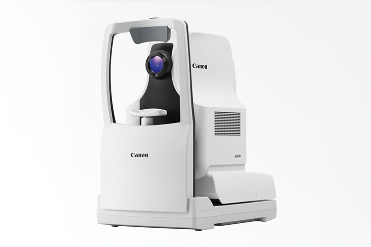
Xephilio OCT-S1: AI-Powered Performance OCTA
Xephilio OCT-S1: AI-Powered Performance OCTA
Xephilio OCT-S1, by Canon Medical, offers superior image quality and a host of automated features to optimize and simplify examinations. It now offers a series of systems with high scanning speed to ensure short examination times, enhance efficiency and improve patient comfort. Our OCTA systems have sophisticated, AI-supported, image postprocessing with 3D projection artefact removal to ensure excellent image quality.
Wide Field Swept Source OCT in One Single Capture
With the Xephilio OCT-S1, Canon Medical introduces revolutionary swept-source technology allowing capture of wide field images of up to 23 mm in a single scan. The Xephilio OCT-S1 enables superior penetration of ocular opacities and provides outstanding tomographic images. It provides a new quality of OCT images in a single scan with greatly reduced noise, increased detail and improved visibility within just seconds.
Swept-source technology allows for a high scanning speed of 100,000 A-scans per second, while maximizing the essential signal data quantity during the acquisition. The 1,060 nm wavelength results in invisible scan lines and therefore ensures better patient collaboration and reduces the impact of the total examination.
Intelligent Denoise
Canon’s RX Intelligent Denoise software was developed using machine learning and an existing clinical OCTA image database to create a neural network for deep learning.
Deep learning neural network (DLNN) artificial intelligence (AI) software integration in eye care has shown notable applications in both image quality improvement and reducing acquisition time. The software decreases the graininess of OCTA images by increasing the signal to noise ratio without loss of anatomical data, therefore, improving diagnostic value within seconds.
CSI Segmentation
With the improved visualization of the choroid layer; the Choroidal Sclera interface can now be detected.
Normative Database
The capabilities of the Xephilio OCT-S1 were extended by comparing a patient's retinal nerve fiber layer (RNFL) and macular thickness measurements to a normative database, clinicians can more accurately identify deviations from normal values, aiding in the early detection of conditions like glaucoma and macular degeneration. The ability to track changes in retinal structures over time is crucial for managing chronic ocular diseases.
Finally, it serves as a reliable reference point, enhancing the precision of measurements and ensuring consistent, reproducible results across different patient populations.
Other blogs that may interest you
Social Media


