


Canon Medical Academy
Vascular Days
Starting April 3
Canon Medical Academy Vascular Days
Starting April 3
Vascular Days
Starting April 3
Webinars on Innovations in Vascular Disease Management and Treatment
Nowadays, partly due to the rising prevalence of obesity and diabetes, vascular diseases are increasingly common in many countries around the world. The most common vascular diseases include peripheral artery disease (PAD), carotid artery disease, and aortic aneurysm disease. Aortic aneurysm ranks globally among the top 10 causes of cardiovascular deaths and is a leading cause of mortality from large vessel diseases. 1 , 2
Canon Medical, together with our partners, strives to bring together experts in the field to share discoveries and insights from research and clinical practice.
The Canon Vascular Days consist of daily webinars (starting at 7 pm CEST for 1h) led by subject matter experts from across the globe. These sessions will provide a platform for professional education as we discuss state-of-the-art imaging and clinical management using illustrative clinical examples.
You can watch the webinars on-demand by clicking on the link below.
Day 1 | Peripheral Artery Disease – Non-Invasive Imaging Ultrasound/CT/MRI
Program
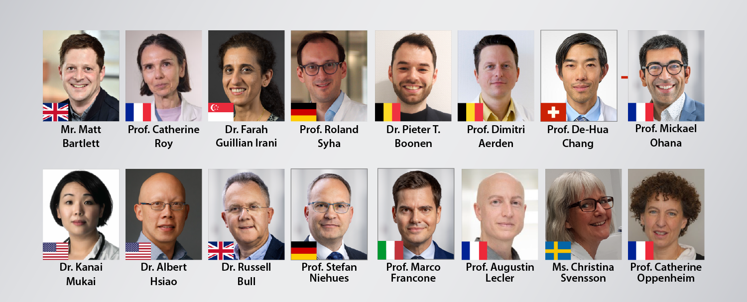
- Lower Limb Doppler Waveform Interpretation in Challenging Cases | Dr. Matt Bartlett
- Advanced CT Imaging in Peripheral Artery Disease | Prof. Catherine Roy
- Clinical Cases in Vascular Imaging | Dr. Kanae Mukai
- Live Q&A | Moderator: Dr. Kanae Mukai
- Identify the global prevalence and mortality trends of Peripheral Artery Disease (PAD) and recognize the disproportionate impact on women compared to men.
- Understand the significance of targeted efforts aimed at reducing PAD risk factors, integrating non-invasive imaging techniques like ultrasound, CT, and MRI.
A systematic review by P. Song et al.3 estimates that peripheral artery disease (PAD) impacts 200 million individuals worldwide, with the global prevalence and mortality rates associated with PAD increasing and affecting women disproportionately compared to men.
There is an urgent need for targeted efforts to both reduce and treat PAD risk factors. To address this, initiatives are already underway to enhance care for PAD patients, specifically targeting high-risk populations with comprehensive, evidence-based preventive care. The widespread adoption and integration of these practices into comprehensive care models help mitigate amputation in the highest-risk populations.
As imaging technology continues to advance, a team of subject matter experts and key opinion leaders will take you through the latest in non-invasive imaging as we work toward continual improvement in the care of patients with PAD.
Dr. Matt Bartlett (Royal Free London NHS Foundation Trust, London, UK)
Prof. Catherine Roy (Strasbourg University Hospital, Strasbourg, France)
Dr. Kanae Mukai (Salinas Valley Health System, Monterey, California, USA)
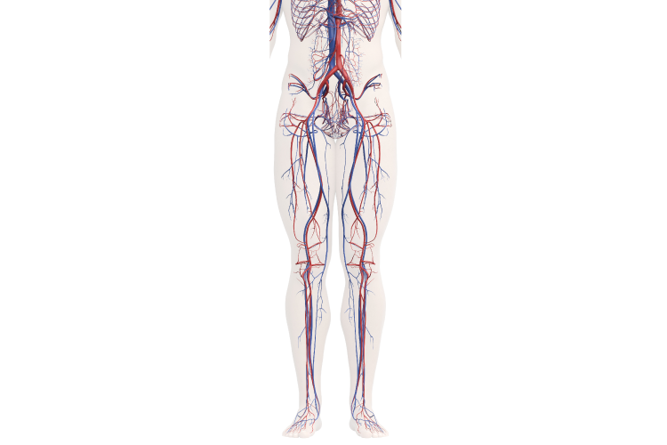
Day 2 | Peripheral Artery Disease – Treatment Interventional Radiology
Program
- The Hybrid Angio-CT Suite: Pushing the boundaries in IR | Dr. Farah Guillian Irani
- Challenges in Peripheral Artery Disease (PAD) – Complex Recanalization Techniques in Chronic Total Occlusion (CTO) | Prof. Roland Syha
- Future prospects of 4D CT in Vascular Surgery | Dr. Pieter Thomas Boonen & Prof. Dimitri Aerden
- Live Q&A | Moderator: Prof. De-Hua Chang
- Understand non-invasive treatments for Peripheral Artery Disease by analyzing case studies on revascularization techniques.
- Recognize challenges in Peripheral Artery Disease and learn intervention strategies.
The global prevalence of PAD continues to grow in parallel with the expanding burden of atherosclerotic risk factors and an aging population. With over 230 million people affected worldwide 4, atherosclerotic lower extremity peripheral artery disease is being increasingly recognized as an important cause of cardiovascular morbidity and mortality.
Traditional cardiovascular risk factors, including advanced age, smoking, and diabetes, are strongly linked to an increased risk of PAD. Although peripheral artery disease has been historically underappreciated compared with coronary artery disease and stroke, the greater attention PAD has received in recent years has led to important new epidemiological insights in the areas of thrombosis, inflammation, dyslipidemia, and microvascular disease.
As treatment pathways continue to evolve, our team will guide you through case studies of revascularization, sharing both the lessons we've learned and the insights we've gained.
Dr. Farah Guillian Irani (Singapore General Hospital, Sengkang General Hospital, SINGHEALTH, Singapore)
Prof. Roland Syha (Stiftungsklinikum Proselis gGmbH Standort: Prosper-Hospital, Recklinghausen, Germany)
Dr. Pieter Thomas Boonen & Prof. Dimitri Aerden (University Hospital Brussels, Brussels, Belgium)
Prof. De-Hua Chang (Luzerner Kantonsspital, Luzern, Zwitserland)
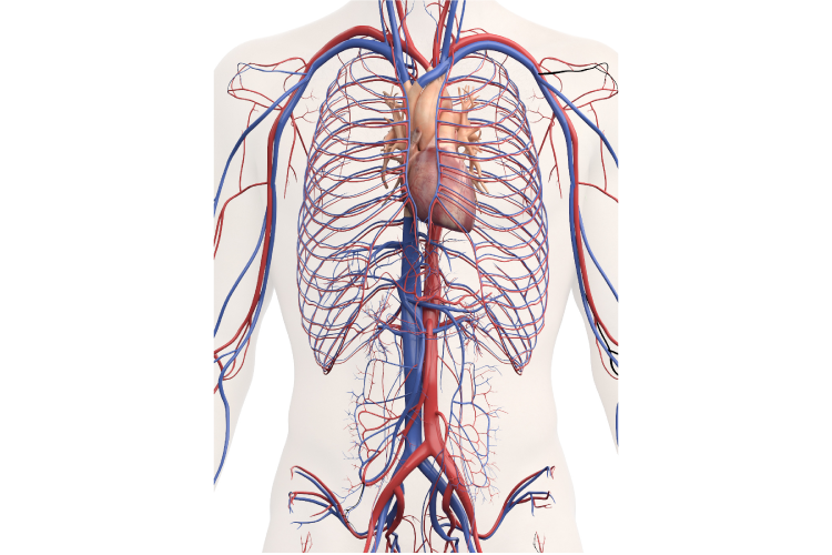
Day 3 | Large Vessel Disease CT/MRI
Program
- EVAR Follow-Up with CT Angiography: Tips and Tricks | Prof. Mickaël Ohana
- Clinical Cases in Vascular Imaging | Dr. Kanae Mukai
- Live Q&A | Moderator: Dr. Albert Hsiao
- Acquire knowledge of modern diagnostic imaging and clinical practices for early detection and effective management of aortic aneurysms, aiming to improve patient outcomes and reduce mortality rates
Aortic aneurysms (AA) caused approximately 160,000 deaths in 2021, making them a leading cause of mortality among large vessel diseases. The number of AA-related deaths is projected to increase by 42%, reaching 244,685 by 2030 (compared to 2019)5. Survival rates closely depend on timely treatment, with recent advances significantly improving outcomes, especially in developed countries.
Despite progress in understanding AA, challenges in early screening and managing aneurysm rupture remain.
Current guidelines emphasize a holistic approach to managing aortic disease, underscoring the importance of modern imaging for comprehensive aorta evaluation.
Our speakers will illustrate the latest in diagnostic imaging techniques and treatments within their clinical practice.
Prof. Mickaël Ohana (Strasbourg University Hospital, Strasbourg, France)
Dr. Kanae Mukai (Salinas Valley Health System, Monterey, California, USA)
Dr. Albert Hsiao (UC San Diego Health, San Diego, USA)
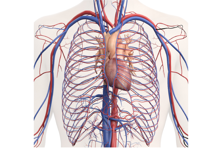
Day 4 | Pulmonary Embolism CT
Program
- CT Perfusion Mapping in Routine Clinical Practice: Optimizing Diagnosis and Care | Dr. Russell Bull
- Imaging and Diagnostic Tools in Pulmonary Embolism CT | Prof. Stefan Niehues
- Live Q&A | Moderator: Prof. Marco Francone
- Assess the significance of imaging techniques in determining PE severity, location, and associated complications.
Pulmonary embolism (PE) is a life-threatening condition resulting from dislodged thrombi occluding the pulmonary vasculature; it can be difficult to diagnose, and it's a serious and prevalent cause of vascular disease.
PE is the third most common cause of cardiovascular death and is associated with multiple inherited and acquired risk factors as well as advanced age.6
Discover the imaging techniques used by our speakers to diagnose and treat pulmonary embolism.
Dr. Russell Bull (Royal Bournemouth Hospital, Bournemouth, UK)
Prof. Stefan Niehues (Caritas Clinics, Berlin, Germany)
Prof. Marco Francone (Humanitas, Milan, Italy)
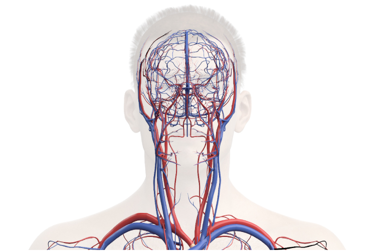
Day 5 | Vasculitis Ultrasound/MRI
Program
- Transforming Arteritis Diagnosis: Ultrasound and SMI – A Non-Invasive Alternative to Biopsies | Christina Svensson
- Cerebral vasculitis: technique and practice | Prof. Augustin Lecler
- Live Q&A | Moderator: Prof. Catherine Oppenheim
- Recognize clinical manifestations and differential diagnoses of vasculitis through case-based learning.
- Improve diagnostic skills for distinguishing vasculitis from other neurological disorders using clinical imaging techniques.
The many forms of vasculitis are characterized by inflammation of blood vessels. Accurate estimation of the incidence and prevalence of vasculitis has been hampered by the absence of reliable diagnostic criteria and the rarity of these conditions. However, much progress has been made over the past two decades, although data from many parts of the world are still lacking.
All forms of cerebral vasculitis are relatively rare, but all are serious and potentially life-threatening. Diagnosing primary central nervous system (CNS) vasculitis is often difficult. There are neither specific clinical features nor a classical clinical course, and no blood or imaging investigations that can confirm the diagnosis. 7, 8
Join the experts as they paint a picture of how they navigate this unique disease.
Christina Svensson (Linköping University Hospital, Linköping, Sweden)
Prof. Augustin Lecler (Hospital Foundation Adolphe de Rothschild, Paris France)
Prof. Catherine Oppenheim (Centre Hospitalier St. Anne, Paris, France)
Join the Canon Medical Academy‘s Webinars
This webinar occurs in the past. The recording will be available soon.
Other webinars that may interest you
References
Disclaimers
Social Media


