Search

Image

CR-10

Full Auto Non-Mydriatic
Retinal Camera
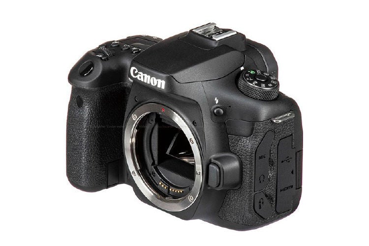
32.5 MP Purpose-built digital EOS camera

Canon is a well-known leading camera manufacturer that has created a unique digital camera called the EOS Retina, which is specifically designed for ophthalmic photography. This camera is integrated with a DIGIC image processor that uses dedicated algorithms to provide optimal image parameters for retinal imaging. As a result, the EOS Retina delivers the best possible retinal image with accurate representation of true colors.

In the CR-10, this camera is now part of the optical system, which further enhances the quality of the retinal images captured. Canon EOS Retina advanced technology being integrated into the latest CR-10 to improve eye care diagnostic capabilities and patient outcomes.

Image

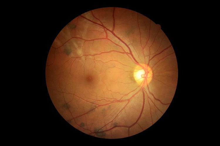
High definition image quality

See more than ever before with the Canon EOS 32.5 MP digital camera and very latest high quality optics.

Image

Extensive Photography Modes

To cover your various imaging requirements.

Image

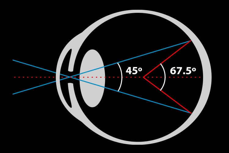
45 degrees images

The imaging standard for retinal screening (67.5 degrees when using center of eye as reference). Additionally a X 2 magnification (30 degrees) is available.

Image

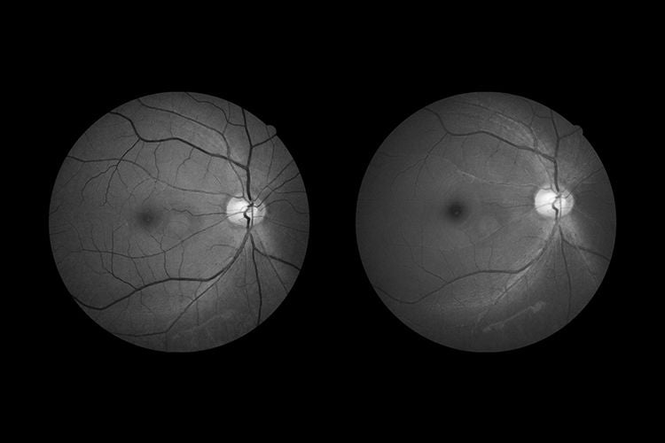
Digital Red Free and Cobalt imaging

The images will be automatically generated from the raw color image data. So no additional capturing of images is required. Canon’s proprietary image processing provides an image quality fully comparable with optical filters.

Image

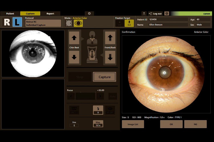
Anterior Photography

Quick and easy anterior segment photography to document the cornea, pupil, eyelid and sclera.

Image

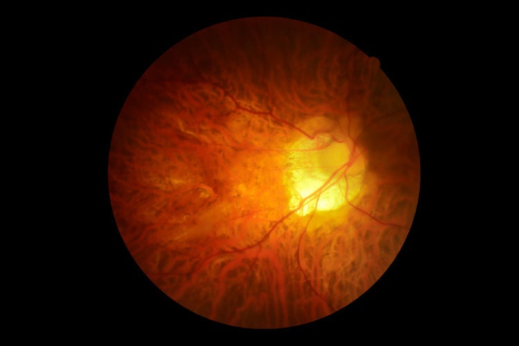
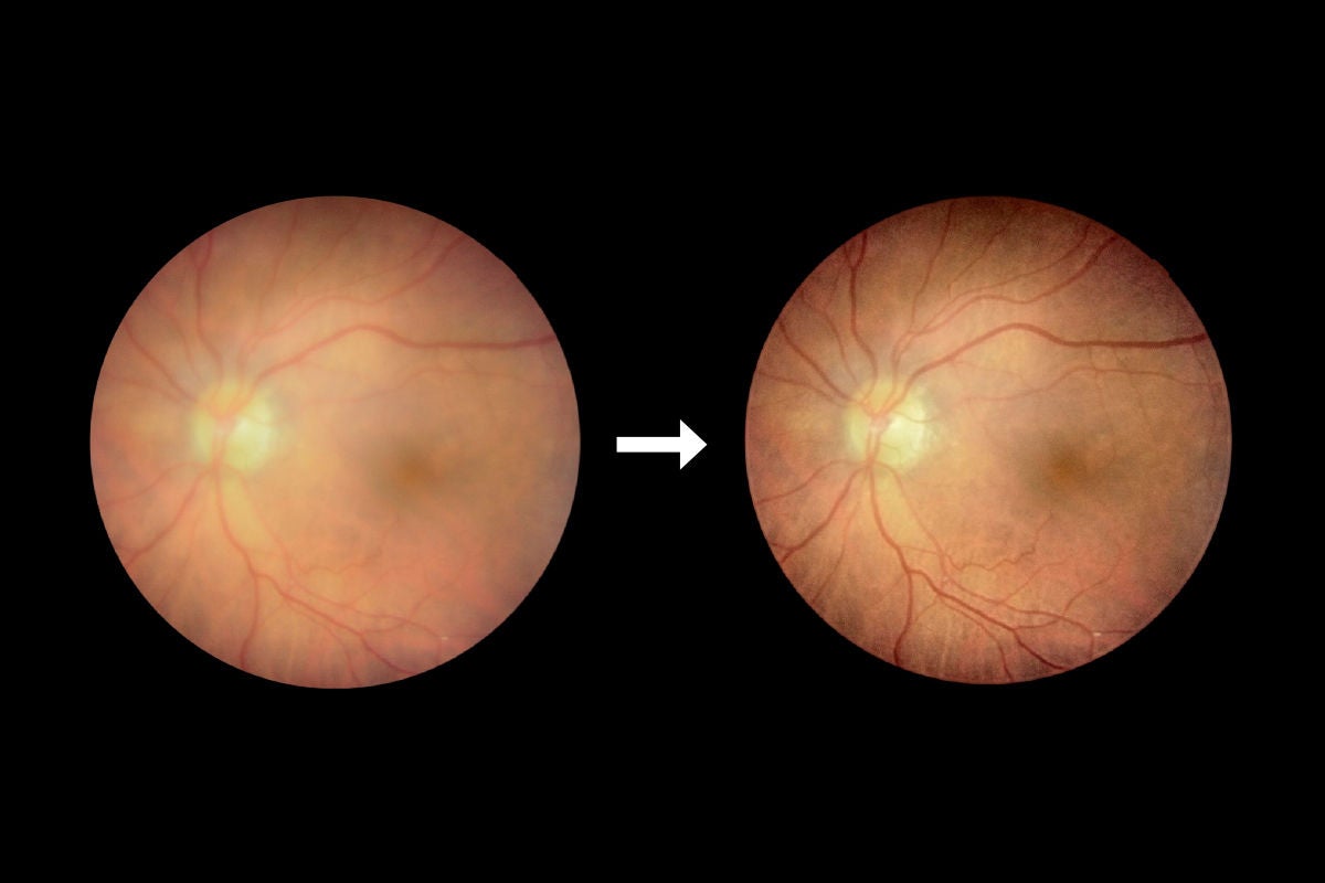
Canon Opacity Suppression

Ocular opacities can obstruct image clarity, by scattering light and blurring the edges of blood vessels, and reducing the difference in brightness of the retina. Furthermore, a cataract can cause images to appear more yellow, thereby hindering the clarity of structures.

With our unique and sophisticated Canon Opacity Suppression software tool, the original brightness and color of the retina will be restored. Our technology is designed to reduce the effect ocular opacity for a clearer view of the blood vessels. This results in more accurate diagnoses as obscure structures become more recognizable for diagnostic purposes.
sou

Retinal Expert software

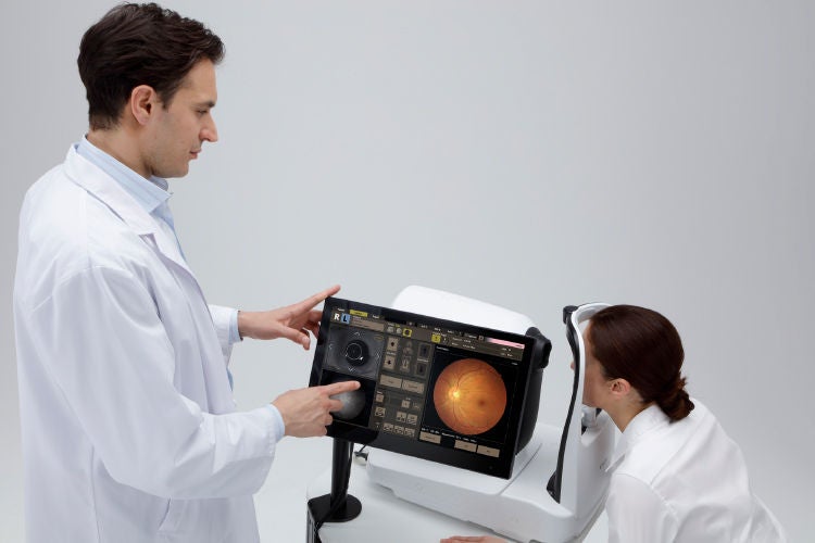
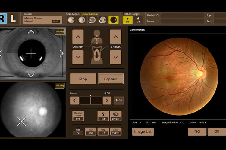
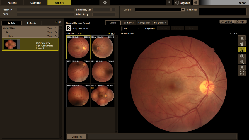
The Graphic User Interface (GUI) of Canon’s RX software is designed to be very intuitive and user-friendly. It is optimized for touch screen operation, with generously dimensioned and clear icons that make navigation effortless.
CR-10

The retinal camera capture screen provides a comprehensive live view of the anterior eye, posterior retina. Large windows allow for easy monitoring of the imaging capture, ensuring precise and accurate results. Even in full auto mode, the RX software offers full control over the CR-10 device, giving the operator the flexibility and assurance they need for an efficient imaging outcome.

Image

Single eye

Offers various ways to display multiple images of the same examination. With the Color/RGB button, the color image also can be displayed in separate RGB channels.

Image

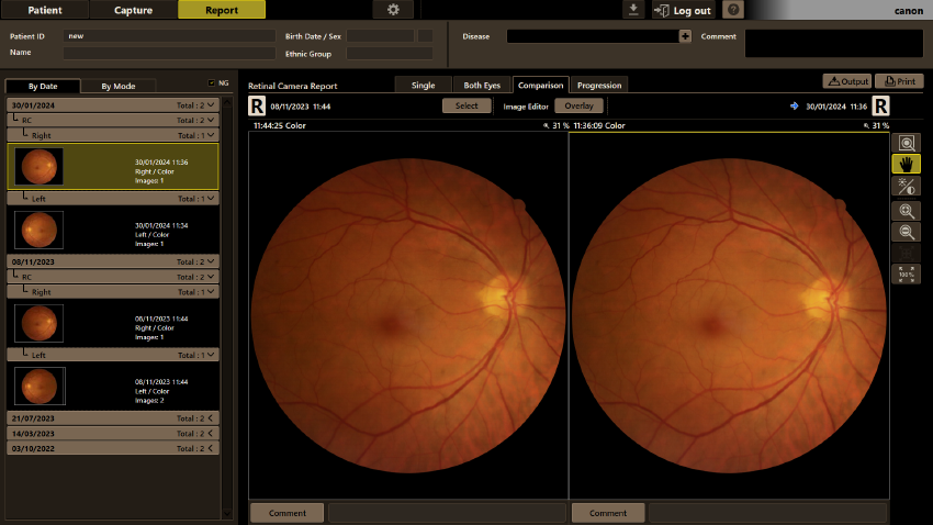
Comparison

Compare result with a previous study. With the overlay functions, changes of the retina can be observed more clearly.

Image

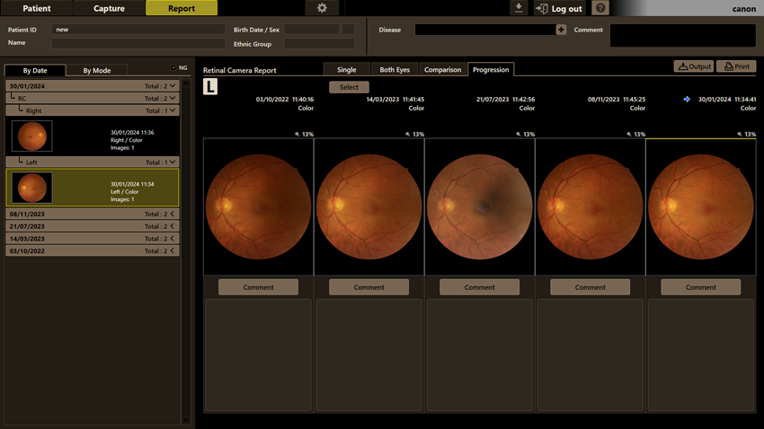
Progression

Select up to five previous studies to observe progression over time.

Image

Efficient Patient Data Management with Retinal Expert Software

The RX software eliminates the need for time-consuming manual input of patient data. It offers the convenience of importing patient lists directly from the practice management system or through a modality worklist in a DICOM environment. This streamlines the workflow and reduces the chance of errors in data entry.

With the cache functionality feature, recent studies are stored on the capture station. This significantly speeds up access to previous examinations as there is no longer a need to wait for downloads due to limited network speed. Clinicians can quickly retrieve and review previous reports, enhancing efficiency and saving valuable time.

RX software seamlessly integrates with your practice software. This means that with just a click, the RX software can automatically open on the specific patient, allowing for easy capturing of new images or reviewing of existing reports. This integration enhances workflow efficiency and ensures a smooth transition between different clinical tasks.

Image

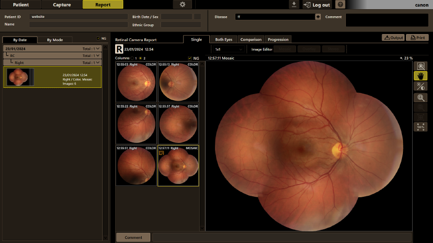
Mosaic

Automatic stitching of up to 20 images for a very wide combined image.

Image

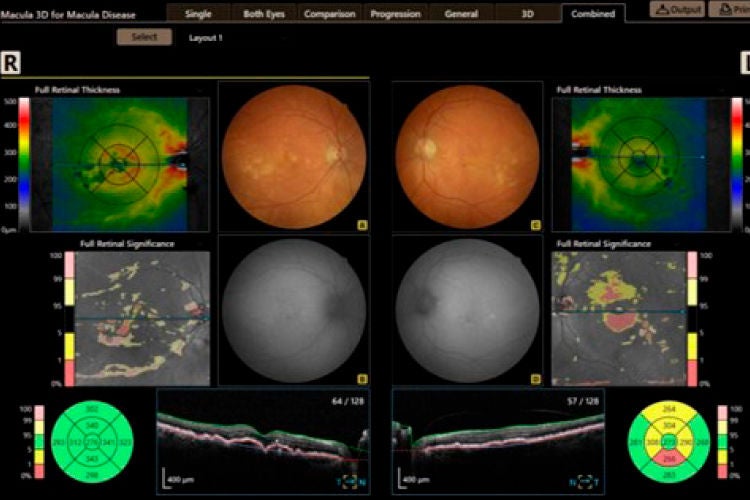
Combined report

Combined analysis results of retinal images, accompanied with OCT scans (from a Canon OCT).

Image

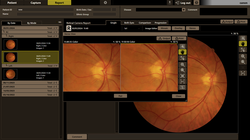
Stereo view

Pairing and viewing 2 images.

Image

Extensive software tools

RX Software offers an impressive arsenal of tools to assist your diagnosis and to create a clear and complete report. Use the emboss function on a retinal image, change its gamma value, adjust its brightness and contrast, change its color balance, add annotations to it, and analyze its C/D ratio. Images can also be rotated, flipped and mirrored.
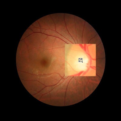
sou Zoom in on pathology.

Loupe function
Zoom in on pathology.
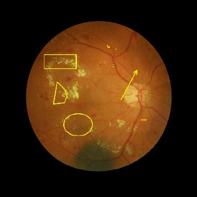
sou Add a shape or texts to a captured image.
Annotations
Add a shape or texts to a captured image.
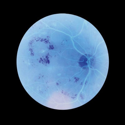
sou Inverts the color of an image to assist diagnosis.

Invert
Inverts the color of an image to assist diagnosis.
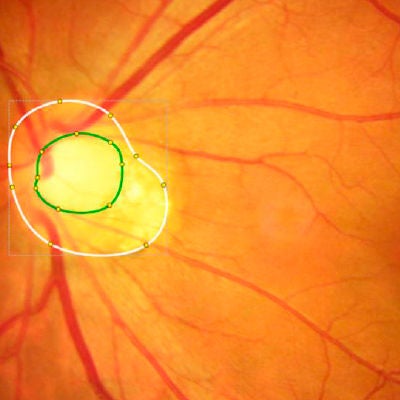
sou Measure the optic nerve papillary area.

Cup/disc measurement
Measure the optic nerve papillary area.
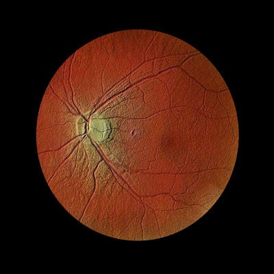
sou The blood vessels stand out.

Emboss Negative
The blood vessels stand out.
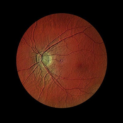
sou The optic disc stands out.

Emboss positive
The optic disc stands out.
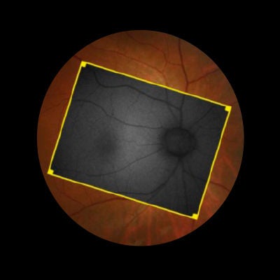
sou Overlay 2 images to see differences and changes in pathology over time.

Overlay
Overlay 2 images to see differences and changes in pathology over time.
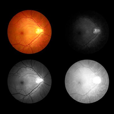
sou View separate RGB channels.

RGB Channel view
View separate RGB channels.