


Clinical gallery
Clinical gallery
Choroidal transplant
70 yo female, autologous choroidal transplant.
Courtesy Dr. Barbara Parolini
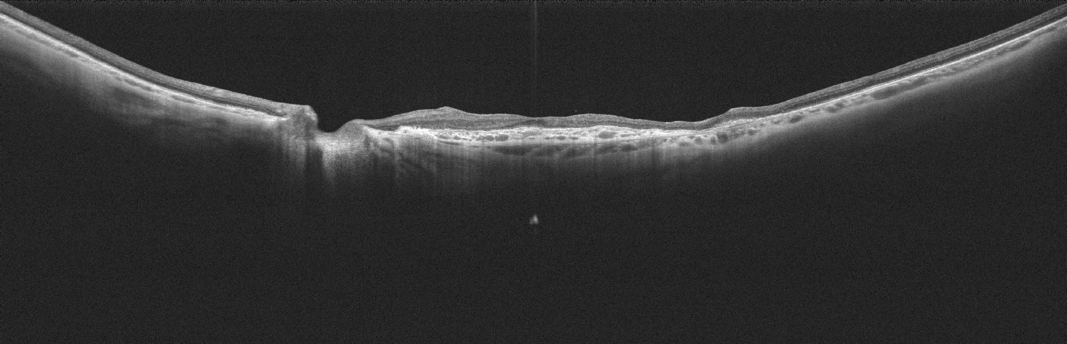 SS-OCT: Choroidal patch can be seen under the fovea.
The Angio-OCT shows the typical and characteristic vascularization of the choroidal patch. The vessels in the patch have different direction compared to the surrounding choroid.
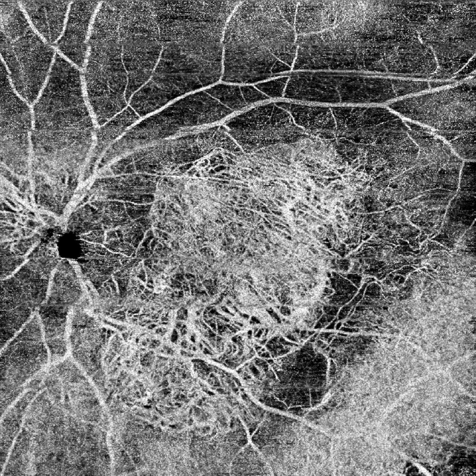 The Angio-OCT shows the typical and characteristic vascularization of the choroidal patch. The vessels in the patch have different direction compared to the surrounding choroid.
Central retinal vein occlusion
Courtesy Dr. Barbara Parolini
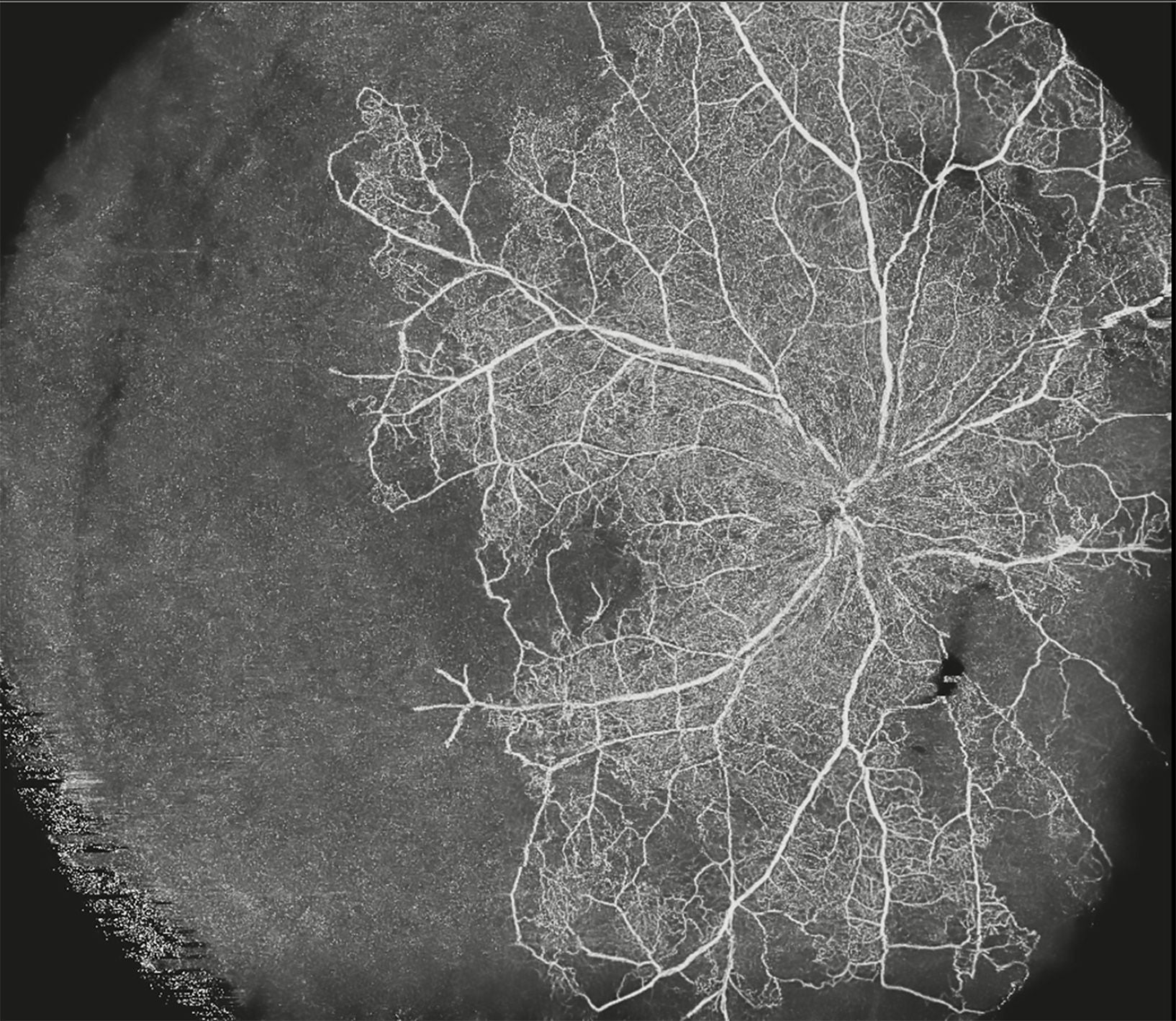 angioOCT of the superficial capillary plexus with Canon OCT-S1. Absence of blood flow in the temporal inferior and nasal quadrants.
angioOCT of the deep capillary plexus with Canon S1. Absence of blood flow in the temporal inferior and nasal quadrants.
OCT Bscan. Thinning of the temporal retina and macular edema of the nasal macula.
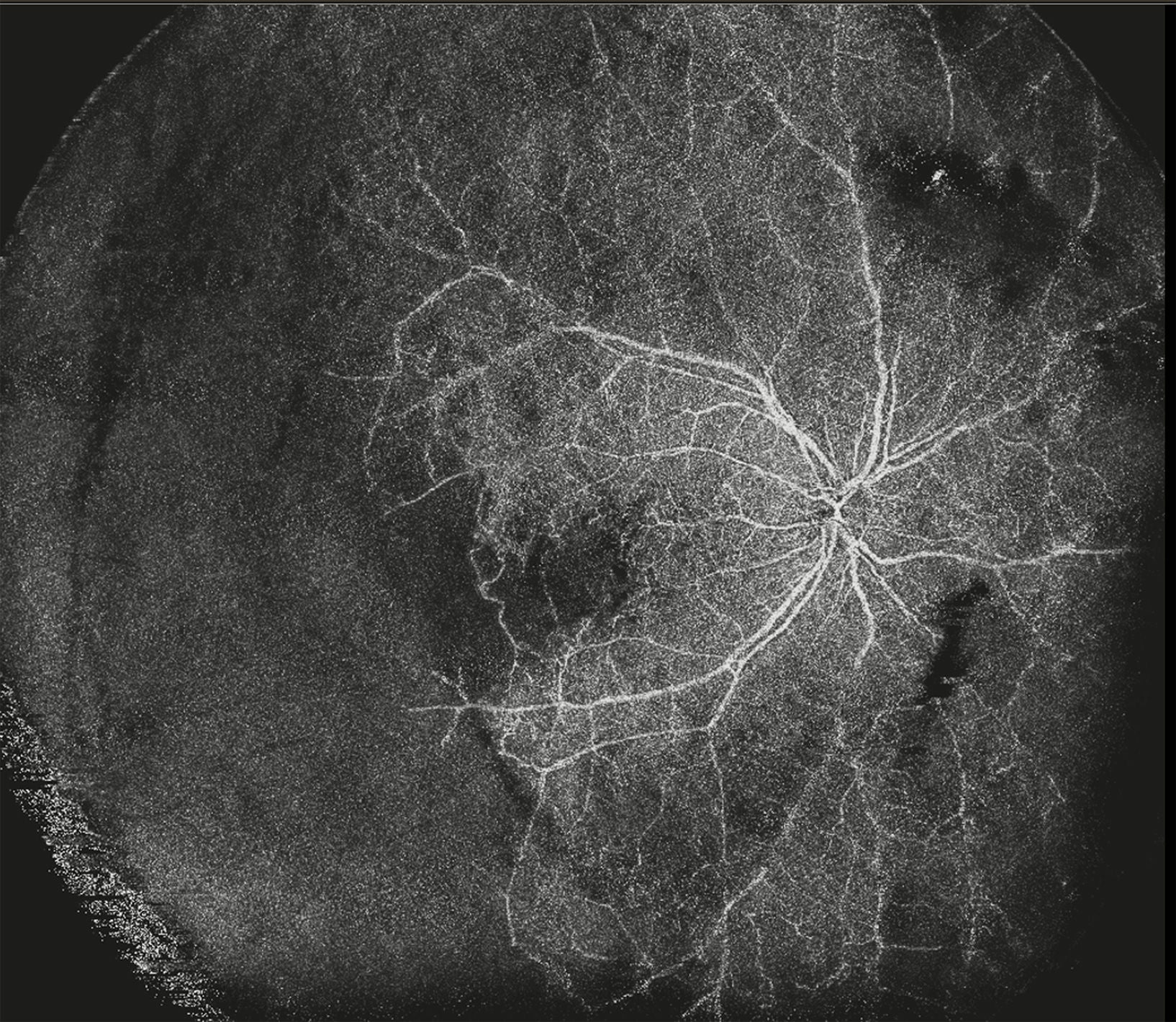 angioOCT of the deep capillary plexus with Canon S1. Absence of blood flow in the temporal inferior and nasal quadrants.
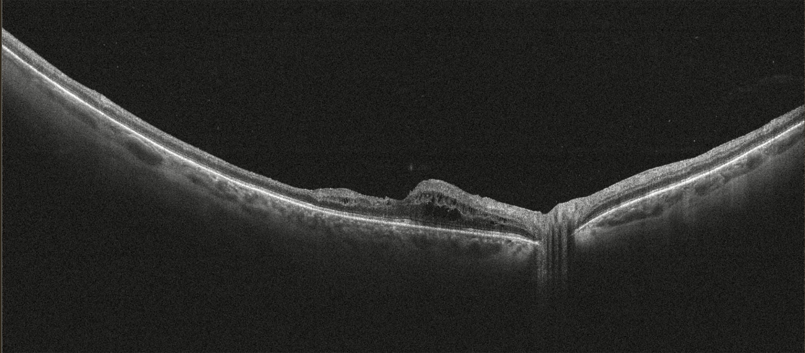 OCT Bscan. Thinning of the temporal retina and macular edema of the nasal macula.
Genetic dystrophy + CME
A 29-year-old patient came to consultation to investigate her genetic disorder further. She has a confirmed diagnosis of a retinal inherited disorder and she already did a genetic test that came out negative. Her VA oscillates between 2/10 and 3/10 in both eyes.
Courtesy Dr. Barbara Parolini
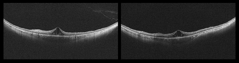 The radial scan shows a Cystoid Macular Edema (CME) and an increased thickness of the choroid (pachychoroid).
Follow up: Patient came to the follow-up consultation, five months after the previous one. She doesn’t report any particular changes in her situation. The left eye is stable. In the right eye, a Lamellar Macular Hole (LMH) is formed from the previous CME. A reduction of the macular thickness is also reported.
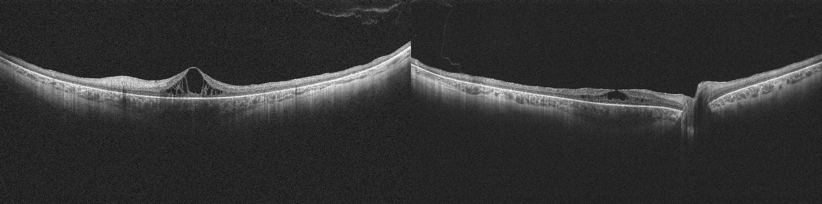 Follow up: Patient came to the follow-up consultation, five months after the previous one. She doesn’t report any particular changes in her situation. The left eye is stable. In the right eye, a Lamellar Macular Hole (LMH) is formed from the previous CME. A reduction of the macular thickness is also reported.
Macula-off retinal detachment
A 55-year-old patient with a history of retinal detachment came to our clinic for a surgery consultation.
Courtesy Dr. Barbara Parolini
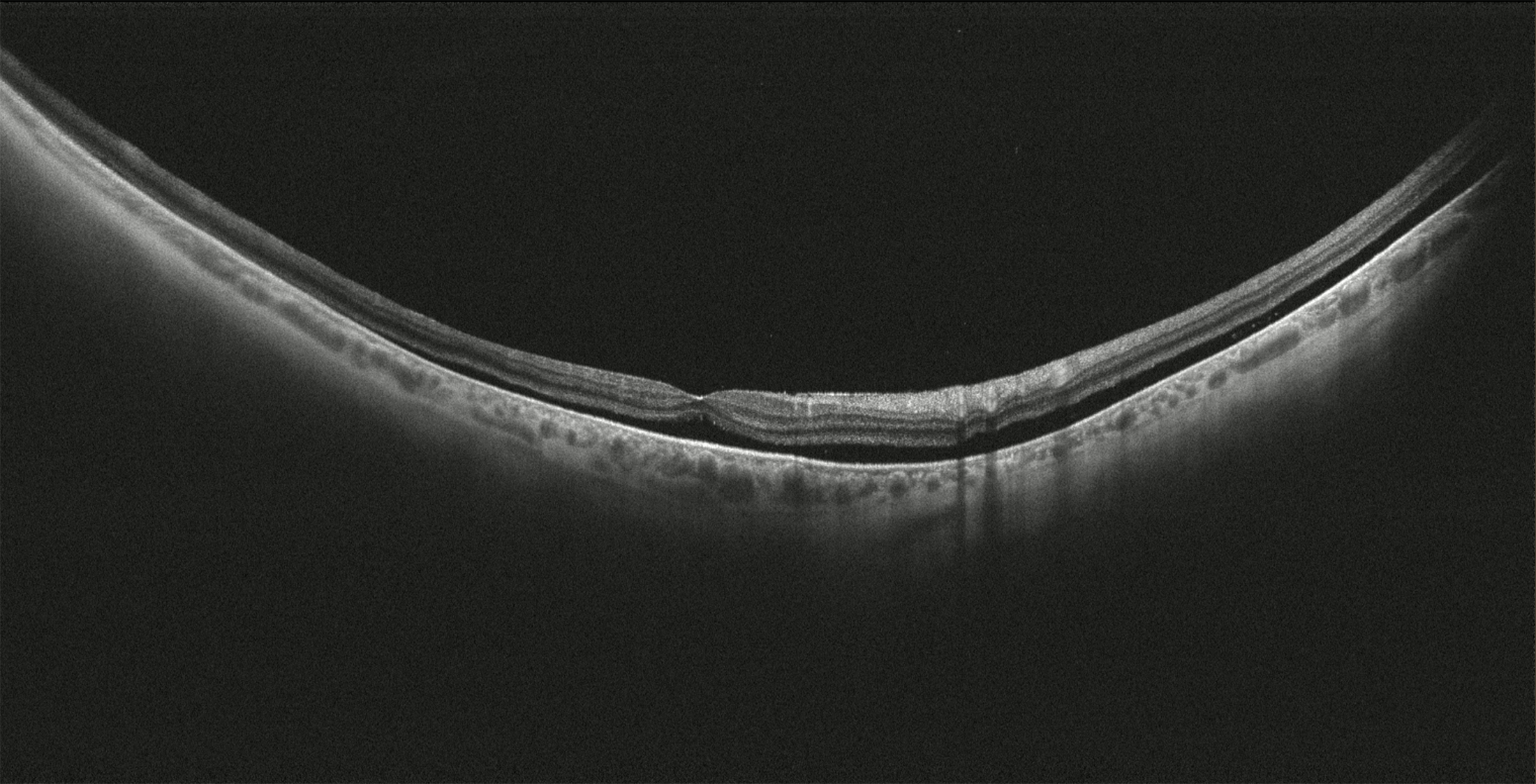 chronic inferior retinal detachment macula-off under silicon oil.
Follow up: Patient came to the first follow-up after the surgery (PPV + silicon oil extraction + removal of the strand + endolaser + silicon oil injection). intraretinal PFCL bubbles are evident.
Follow up: Patient came to the follow-up after the second surgery. Irregular retinal profile but attached. Some really small PFCL bubbles are visible extrafoveal consultation.
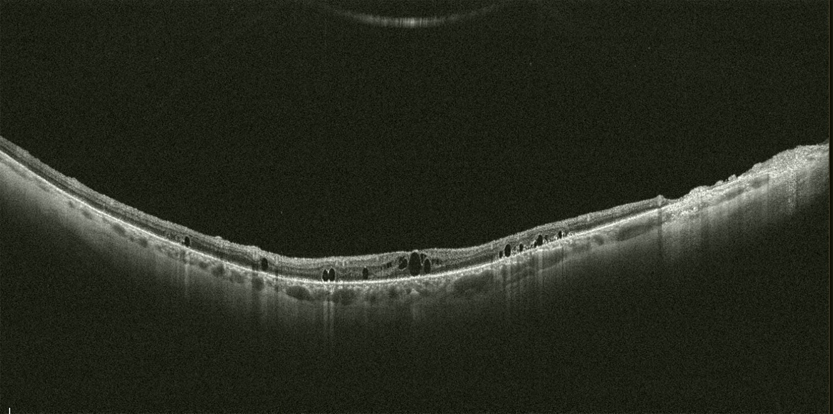 Follow up: Patient came to the first follow-up after the surgery (PPV + silicon oil extraction + removal of the strand + endolaser + silicon oil injection). intraretinal PFCL bubbles are evident.
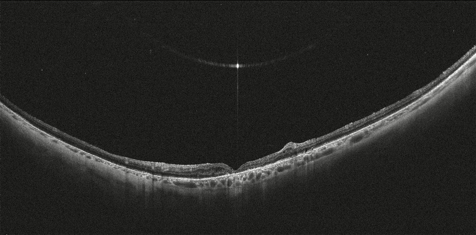 Follow up: Patient came to the follow-up after the second surgery. Irregular retinal profile but attached. Some really small PFCL bubbles are visible extrafoveal consultation.
Trauma with ball
8 y.o. healthy boy, trauma with tennis ball.
Courtesy Dr. Barbara Parolini
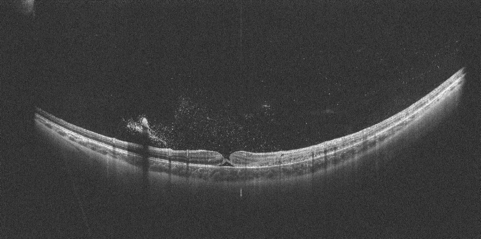 Blood in the vitreous chamber. Partial thickness macular hole with externa; layers still continuous.
Thanks to wide-field OCT scans we can study the retina beneath the vitreous haemorrhage.
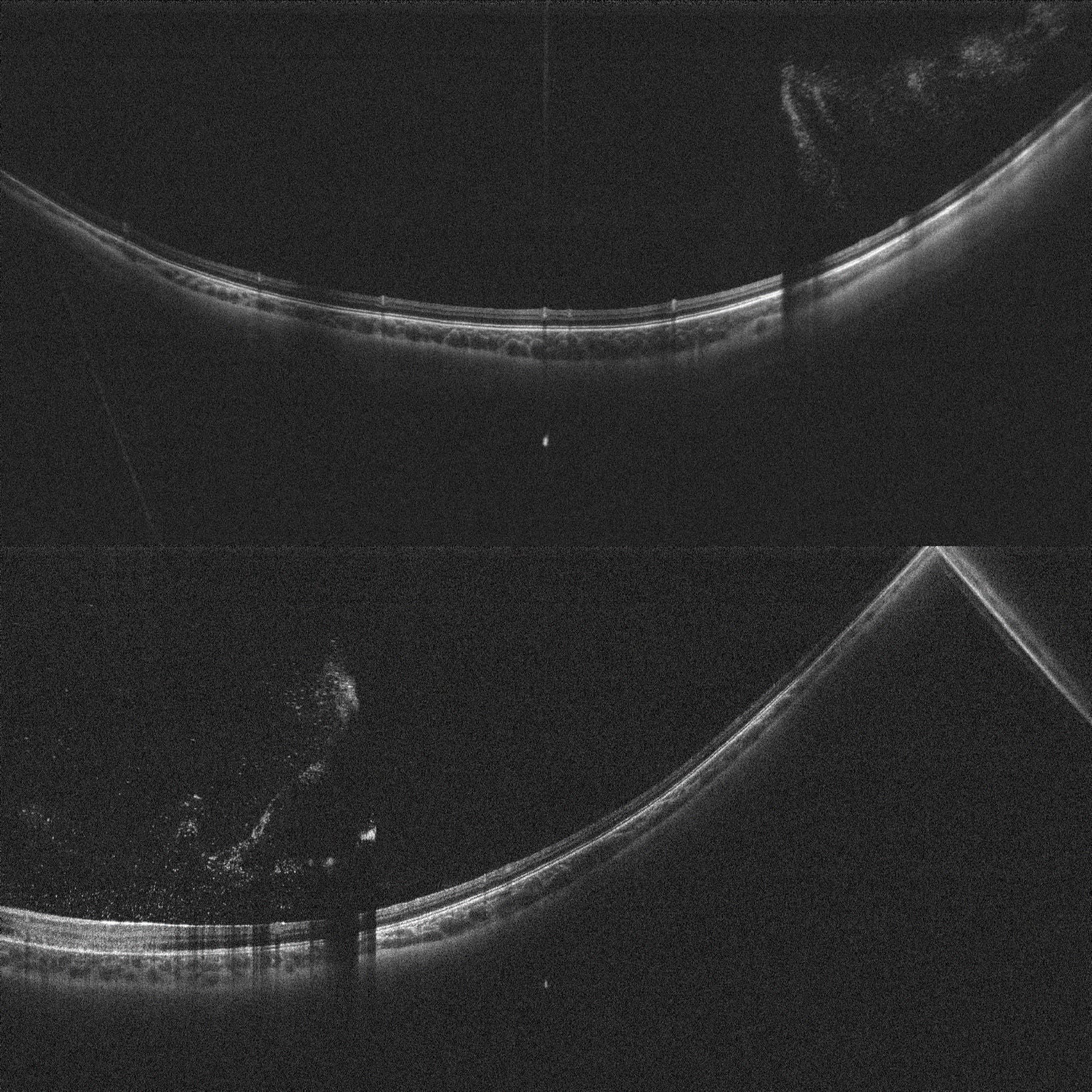 Thanks to wide-field OCT scans we can study the retina beneath the vitreous haemorrhage.
Retinoschisis
Courtesy Prof. Paulo E. Stanga MD
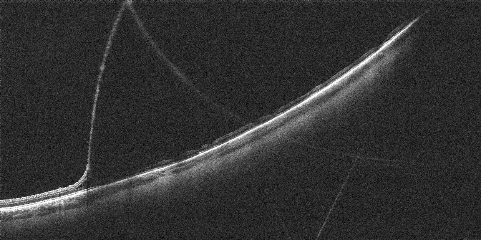 Near infra-red image of the area scanned and scan pattern with cross sectional mid and peripheral single OCT scan showing the inner and outer leaf.
3D OCT rendering showing the retinal inner leaf Peau D’Orange (white arrow) and near infra-red image of the area scanned and scan pattern.
Proliferative diabetic retinopathy
Courtesy Prof. Paulo E. Stanga MD
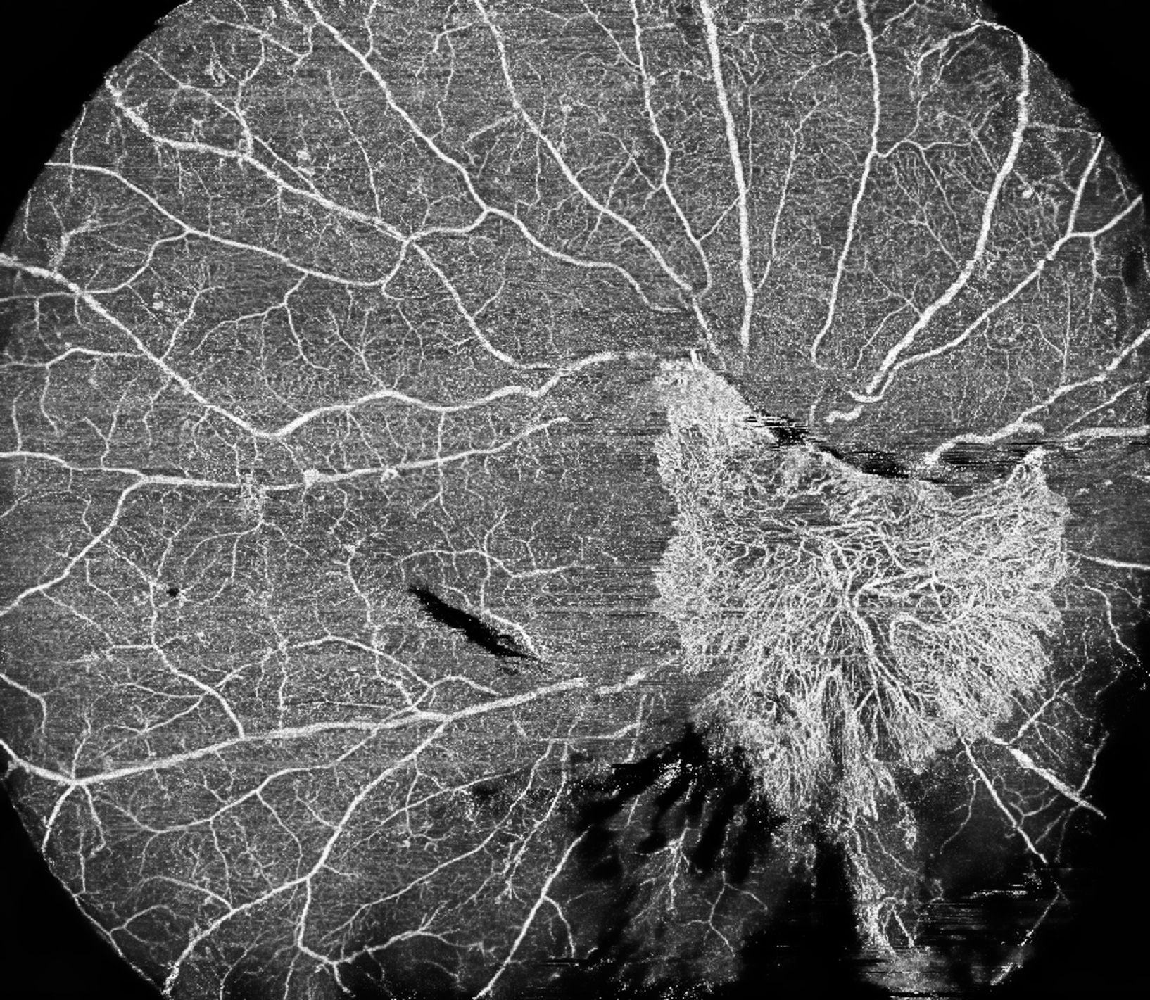 Proliferative Diabetic Retinopathy. Large Frond of Neovascularisation at the Disc (NVD) with Mid-Periheral Ischaemia on OCT-A.
Cross-sectional scans and 3-D Renderings showing NVD, “Tabletop” Posterior Vitreous Detachment (PVD), Tractional Schisis on points of Vitreoretinal Adhesion, Posterior Pole Epiretinal Gliosis/ERM, Intraretinal Fluid/Diabetic Macular oedema (DMO) and Intravitreal red Blood Cells (RBC).
Proliferative diabetic retinopathy
Bilateral Proliferative Diabetic Retinopathy with Diabetic macular oedema. Treated elsewhere with Retinal Laser and with a course of Intravitreal anti-VEGF Injections.
Courtesy Prof. Paulo E. Stanga MD
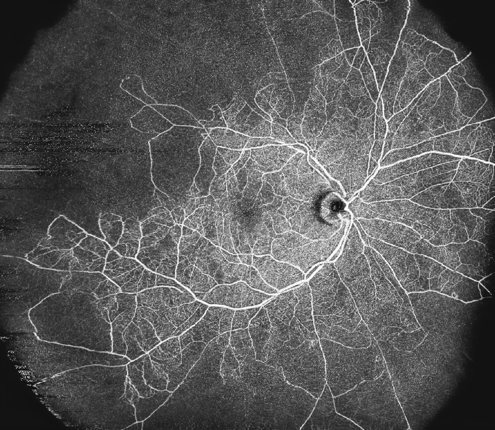 OCT-A clearly shows Mid and Peripheral Non-Perfusion (Ischaemia) with “Pruning” of the Foveal Avascular Zone (FAZ) in the Right Eye and a well-perfused Fovea in the Left Eye, in a non-invasive manner. OCT-A ruled out any Neovascularization Elsewhere (NVE).
OCT-A clearly shows Mid and Peripheral Non-Perfusion (Ischaemia) with “Pruning” of the Foveal Avascular Zone (FAZ) in the Right Eye and a well-perfused Fovea in the Left Eye, in a non-invasive manner. OCT-A ruled out any Neovascularization Elsewhere (NVE).
Cross-sectional scans show the presence of Intraretinal Fluid/DMO and ruled out the presence of a PVD, though a cleavage plane between the posterior cortical vitreous and the neuroretina is already visible, and the left eye already shows a weiss ring in the process of detaching form the ONH.
Cross-sectional scans show the presence of Intraretinal Fluid/DMO and ruled out the presence of a PVD, though a cleavage plane between the Posterior Cortical Vitreous and the Neuroretina is already visible, and the Left Eye already shows a Weiss Ring in the process of detaching form the ONH.
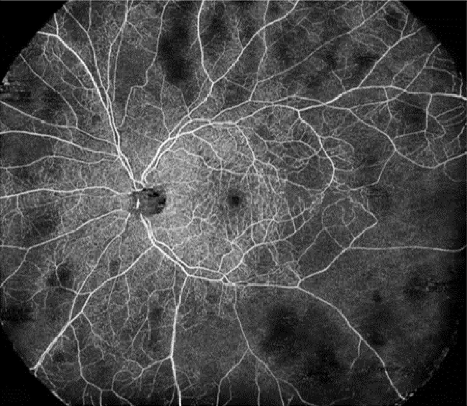 OCT-A clearly shows Mid and Peripheral Non-Perfusion (Ischaemia) with “Pruning” of the Foveal Avascular Zone (FAZ) in the Right Eye and a well-perfused Fovea in the Left Eye, in a non-invasive manner. OCT-A ruled out any Neovascularization Elsewhere (NVE).
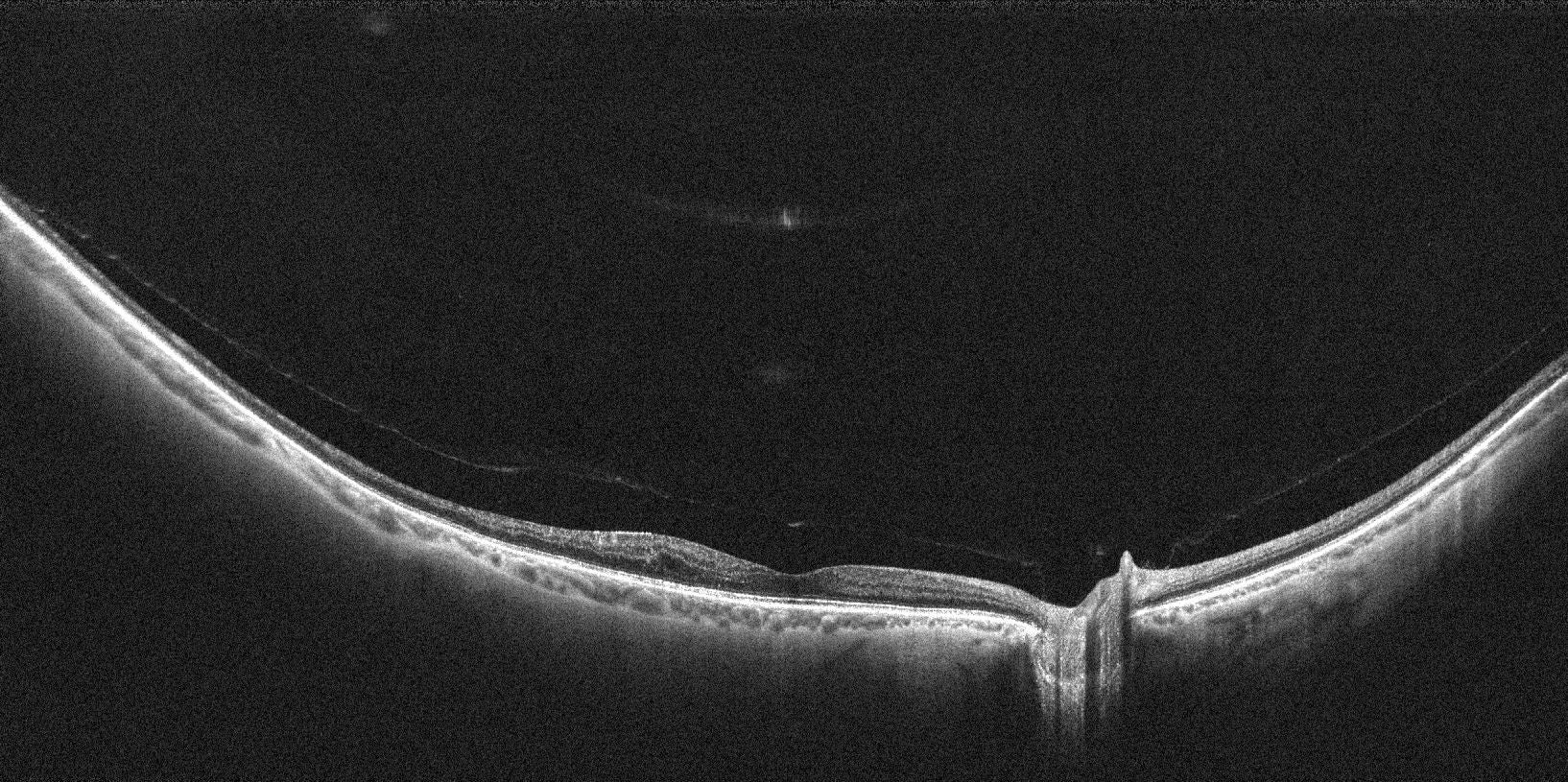 Cross-sectional scans show the presence of Intraretinal Fluid/DMO and ruled out the presence of a PVD, though a cleavage plane between the posterior cortical vitreous and the neuroretina is already visible, and the left eye already shows a weiss ring in the process of detaching form the ONH.
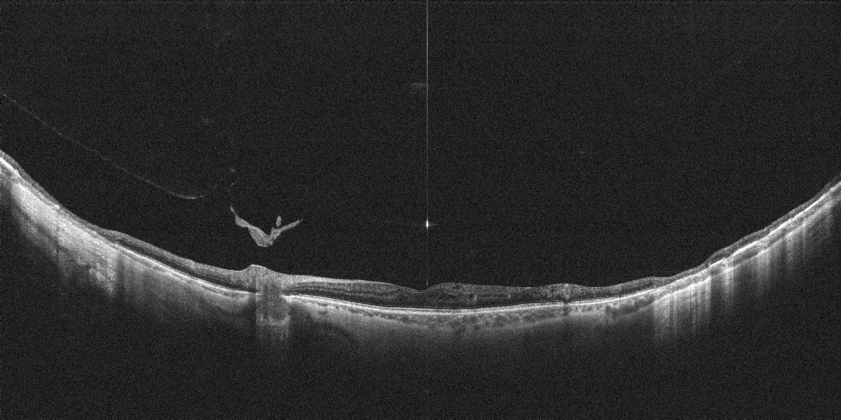 Cross-sectional scans show the presence of Intraretinal Fluid/DMO and ruled out the presence of a PVD, though a cleavage plane between the Posterior Cortical Vitreous and the Neuroretina is already visible, and the Left Eye already shows a Weiss Ring in the process of detaching form the ONH.
OS tear at the edge of the lattice, followed by retinopexy
Courtesy Prof. Paulo E. Stanga MD
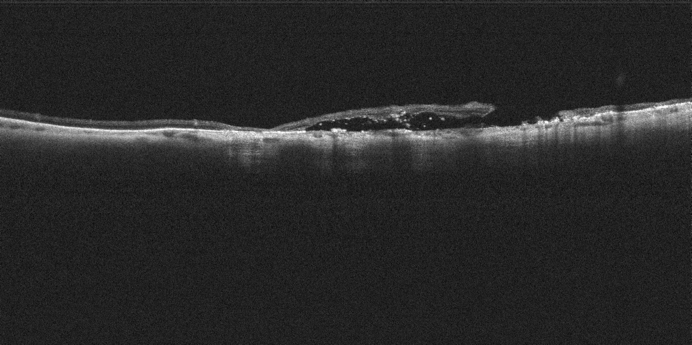 Cross-Sectional scan confirmed the presence of a PVD and a normal macula.
3-D rendering shows the elevated flap of the tear and revealed the vitreous attachment to it.
Asteroid Hyalosis and PVD
Courtesy Prof. Paulo E. Stanga MD
Cross sectional scans shows the presence asteroid hyalosis in the absence of PVD in the left eye. Macula is normal.
OS vitreous floaters, peripheral areas of vitreous retinal traction (VRT) and elevated retina plus tears in lattice with VRT and SRF
Courtesy Prof. Paulo E. Stanga MD
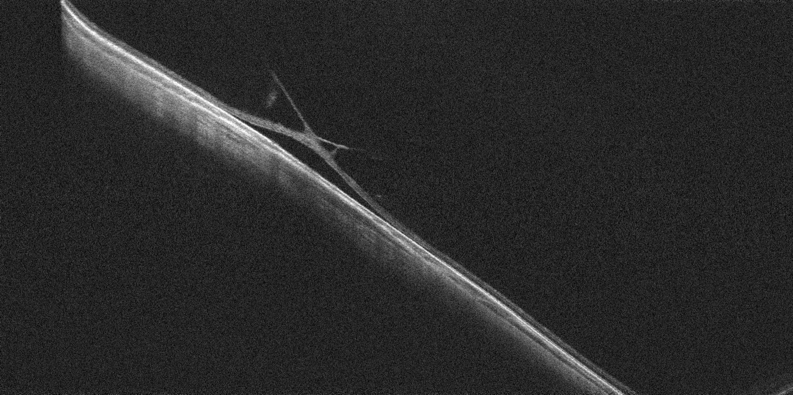 Navigated Peripheral Cross-Sectional OCT scans confirmed the presence of retinal tears when not clear on ophthalmoscopy and revealed the presence of an elevated retina with subretinal fluid/subclinical retinal detachment (RD) and vitreous attachment/traction, possibly indicating the high risk of progression.
3-D Rendering shows increased collagen deposition/vitreous attachment over the area of retinal lattice and area of elevated retina/RD and retinal flap/tear.
OD Tuft
Courtesy Prof. Paulo E. Stanga MD
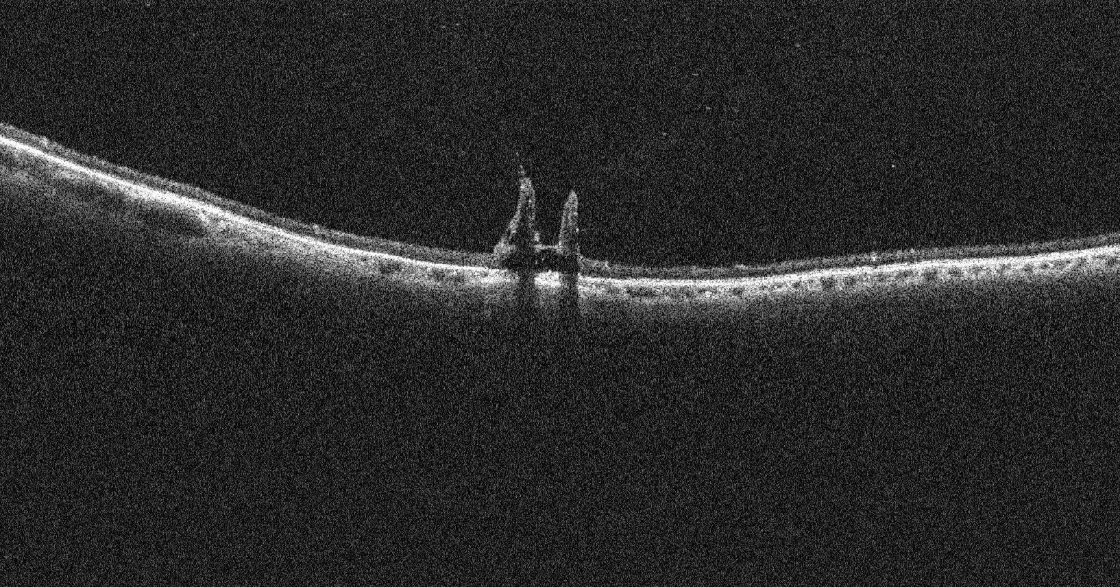 The Cross-Sectional scans revealed an attached vitreous. Inferiorly, a round area of one-disc diameter and compatible with pre-retinal fibrosis was noted. Navigated Peripheral OCT scans showed a visually striking and elevated lesion compatible with a large vitreoretinal tuft. Note the absence of retinal hole/tear and the presence of a vitreous attachment.
OS PVD with vitreous RBC
Courtesy Prof. Paulo E. Stanga MD
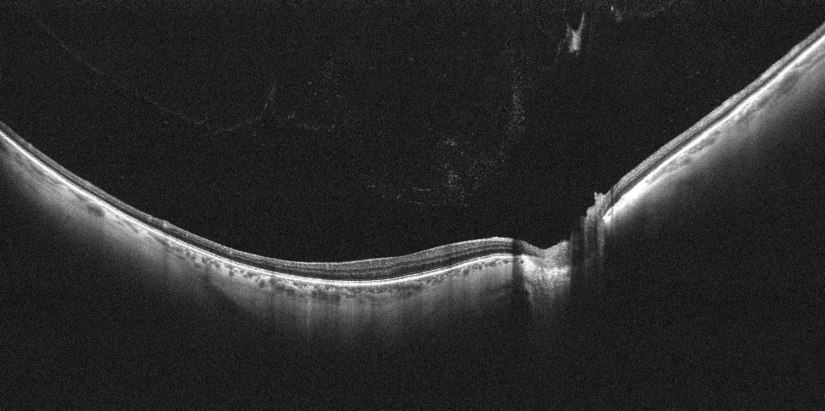 Cross-sectional Scans confirmed the presence of a PVD, showed a Weiss Ring and RBC in the Posterior Cortical Vitreous.
3-D Rendering showing PVD and an enhanced hyper-reflective Posterior Cortical Vitreous due to the presence of RBC.
OS Coloboma
Courtesy Prof. Paulo E. Stanga MD
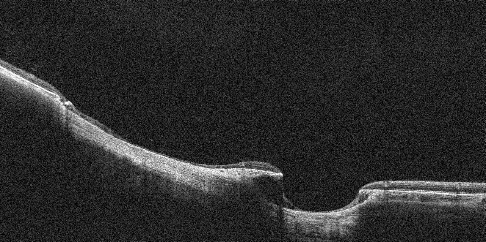 Cross-Sectional scans show the colobomatous depression, the presence of an attached vitreous and ruled out any peri-Coloboma RD.
The 3-D Renderings highlight the findings even further.
OD Bionic Eye with Retinitis Pigmentosa
Courtesy Prof. Paulo E. Stanga MD
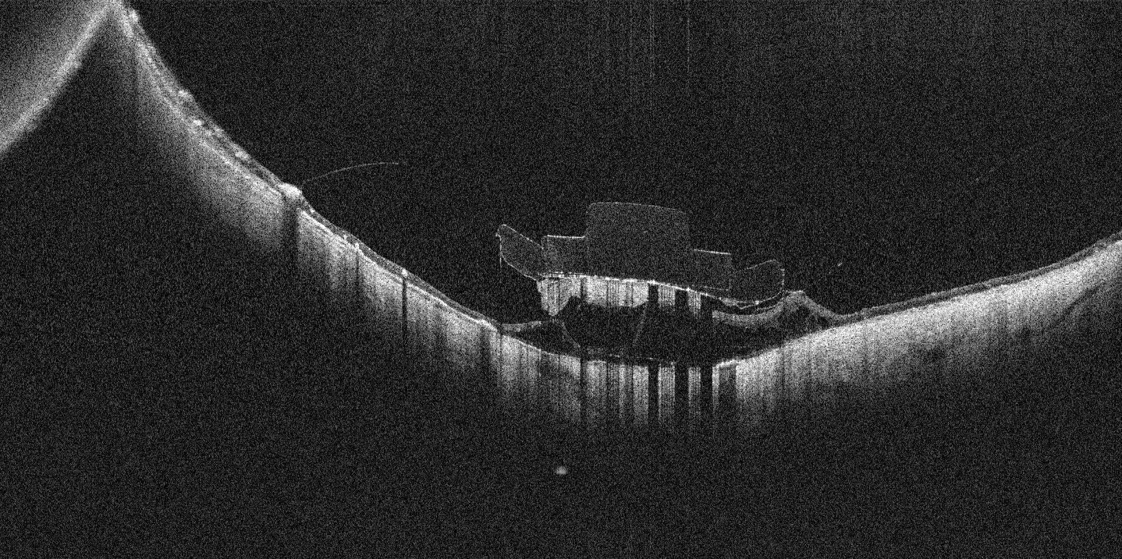 Argus II® Electronic Epi-Retinal Implant a patient with Advanced Retinitis Pigmentosa, implanted by PES 9 years prior to these scans being obtained. Cross-Sectional Widefield OCT scans show the presence of sub-array fibrosis, a gap between the array and the neuroretina, degenerative intraretinal changes and remnants of detached internal limiting membrane (ILM).
Social Media


