Search

Ophthalmology

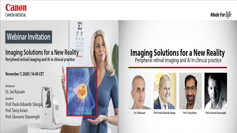
Imaging Solutions for a New Reality: Peripheral retinal imaging and AI in clinical practice

  • Dr. Sal Rassam , Worthing, UK
  • Prof. Paulo Eduardo Stanga , Consultant Ophthalmologist & Vitreoretinal Surgeon, London, UK
  • Prof. Tariq Aslam , Professor of Ophthalmology and Interface Technologies, University of Manchester, Manchester, UK
  • Prof. Giovanni Staurenghi , Chairman University Eye Clinic, Milan, Italy
Image

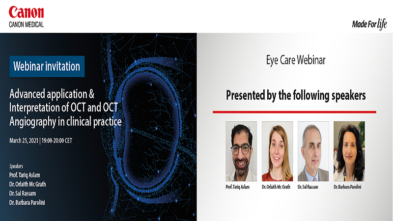
Advanced application & Interpretation of OCT and OCT Angiography in clinical practice

Learning Objectives


  1. The aim of this event is to help ophthalmologists understand and learn how to use OCT and OCTA in daily clinical practice for certain diseases.
  2. Get practical tips from the examples discussed to increase your diagnostic confidence.
  3. In the question-and-answer session, you could find answers that you need.
Image

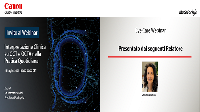
Interpretazione clinica su OCT e OCTA nella pratica quotidiana

Programma


  • Utilità dell'OCT e Angio-OCT Wide Field nella pratica clinica; Dr. Barbara Parolini
  • Live Q&A
  • Italian
Image

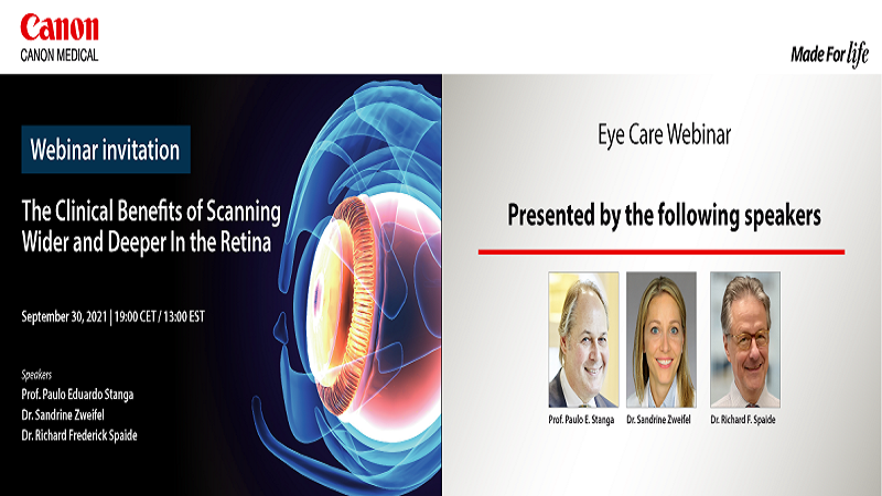
The Clinical Benefits of Scanning Wider and Deeper in the Retina

Program


  • Cross-Sectional, 3D and Topographic Wide field Optical Coherence Tomography (OCT) Imaging of the Peripheral Retina and Vitreoretinal Interface and Assessment of the status of the Posterior Cortical Vitreous - Prof. Paulo Eduardo Stanga
  • Feasibility and Clinical Utility of UWF OCTA - Dr. Sandrine Zweifel
  • Overview of Venous Overload Choroidopathy - Dr. Richard Frederick Spaide
  • Live Q&A
Image

Glaucoma & Wide Field imaging: Learn from the Experts and Gain New Insights!

Program


  • Het gebruik van de OCT bij glaucoom - Dr. Hans. G. Lemij
  • Wide Field OCT en OCT angiografie - Dr. Rogier Müskens,
  • Live Q&A
  • Dutch
Image

Enhance Your Daily Patient Management with Wide Field OCT

Program


  • OCT-A as a window to COVID-19 - the potential – Prof. Tariq Aslam & Dr. Orlaith McGrath
  • Overview of choroidal diseases using the wide field OCT-S1 Xephilio and focus on the pachychoroid disease spectrum. - Dr. Prithvi Ramtohul
  • Objective Assessment of Posterior Vitreous Detachment (PVD) Status and the Peripheral Vitreoretinal Interface & Retina: New 3D 23 mm Wide Field OCT.- Prof. Paulo E. Stanga
  • Live Q&A
Image

What Can Wide Field Imaging Do for Your Practice: Case Studies

Programme


  • Imagerie Moderne du Vitré en OCT – Mr. Adil El Maftouhi
  • La Place de l’OCT et OCTA à Ultra-Grand Champ dans les Pathologies Rétiniennes – Dr. Ali Erginay
  • L'OCTA Grand Champ Peut-Il Remplacer L'angiographie à la Fluorescéine pour la Rétinopathie Diabétique? – Prof. Dr. Ramin Tadayoni
  • Live Q&A
  • French
Image

Disclaimers

  • The opinions expressed in this material are solely those of the presenter and not necessarily those of Canon Medical Systems. Canon Medical Systems does not guarantee the accuracy or reliability of the information provided herein.
  • The clinical results, performance and views described in this video/webinar are the experience of the clinician. Results may vary due to clinical setting, patient presentation and other factors. Many factors could cause the actual results and performance of Canon Medical’s products to be materially different from any of the aforementioned.
  • Some products shown might not be available in all regulatory jurisdictions, please consult with your local Canon sales office for availability in your region
  • Our webinars are organized and provided at no cost. In no circumstance would you be requested to share your bank details or make a payment. Neither will you be asked to share personal/sensitive information. Any correspondence from Canon Medical Systems Europe will include the domain eu.medical.canon. If you are at all unsure or suspect impersonation or any illegitimate communication please contact us through https://eu.medical.canon/contact immediately.