


Innovative Approaches to Diagnosing and Treating Endometriosis and Adenomyosis
Endometriosis and adenomyosis affect an estimated 190 million women worldwide 1 ; nevertheless, the impact of these conditions on quality of life and well-being has long been underestimated.
On average, it takes 7.5 years to diagnose endometriosis or adenomyosis 2 , and even then, patients often struggle to access effective treatment plans. These conditions not only contribute to infertility but also cause chronic pain that can severely diminish a woman's daily life.
Fortunately, awareness and understanding of endometriosis and adenomyosis are growing, leading to advancements in diagnosis and treatment. Today, ultrasound is recognized as a reliable, non-invasive tool for diagnosis, while MRI plays a critical role in identifying and visualizing lesions, adhesions, and the extent of endometrial tissue infiltration, particularly in cases of deep infiltrating endometriosis – often without the need for contrast agents.
In this webinar, we will explore the latest imaging techniques and intervention methods, focusing on how ultrasound and MRI can improve patient outcomes and offer a more compassionate approach to managing these challenging conditions.
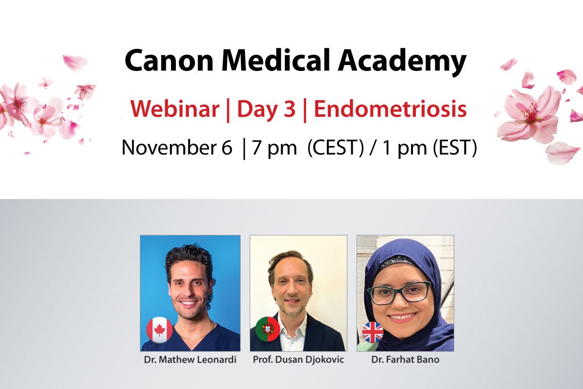
Day 3 | Endometriosis
Ultrasound / MRI
November 6, 7 pm (CET) / 1 pm (EST)
Join us for an insightful webinar on innovative approaches to diagnosing and treating endometriosis and adenomyosis, moderated by Dr. Mathew Leonardi . Featuring expert presentations by Prof. Dusan Djokovic and Dr. Farhat Bano , this session will highlight the latest advancements in ultrasound and MRI techniques to improve patient outcomes.
Register today and be part of this essential discussion!
Program
- Introduction | Dr. Mathew Leonardi
- Ultrasound Mapping of Pelvic Endometriosis in Combination with the #Enzian Classification | Prof. Dusan Djokovic
- Role of MRI in Imaging of Endometriosis | Dr. Farhat Bano
- Live Q&A, moderated by Dr. Mathew Leonardi
- Learn to perform systematic ultrasound assessments for endometriosis, apply the #Enzian classification, and use structured reporting to enhance early diagnosis
- Understand the role of MRI in diagnosing and assessing the extent of endometriosis
- Participating in the live Q&A to find answers to your own questions
Certificate*
All delegates will receive a certificate of attendance.
Speakers and Presentations
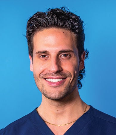
Moderator: Dr. Mathew Leonardi, MD, PhD, FRCSC Assistant Professor
McMaster University Medical Centre
Hamilton, Canada
McMaster University Medical Centre
Hamilton, Canada

Speaker: Prof. Dusan Djokovic, MD, PhD Consultant at Hospital CUF Descobertas
Hospital CUF Descobertas
Lisbon, Portugal
Presentation: Ultrasound Mapping of Pelvic Endometriosis in Combination with the #Enzian Classification
Ultrasound represents the first-line imaging tool in patients with symptoms and signs of endometriosis. When used by experienced operators, its sensitivity and specificity for deep endometriosis may exceed 80 – 90%, although the accuracy varies for different pelvic structures. To reduce the often years-long diagnostic delay in endometriotic patients, the European Society for Human Reproduction and Embryology currently recommends diagnostic laparoscopy only if imaging results are negative and/or empirical treatment is unsuccessful or inadequate.
This presentation promotes the practice of standardized, comprehensive and systematic ultrasound assessment of pelvic endometriosis, in correlation with the #Enzian classification that is a valid and suitable system applicable in both non-invasive and surgical procedures. Recommendations for structured reporting of findings obtained by dynamic ultrasound evaluation will also be presented. Finally, an early diagnosis algorithm will be provided to guide physicians in cases of suspected endometriosis, including basic and expert phases of ultrasound assessment.
Hospital CUF Descobertas
Lisbon, Portugal
Presentation: Ultrasound Mapping of Pelvic Endometriosis in Combination with the #Enzian Classification
Ultrasound represents the first-line imaging tool in patients with symptoms and signs of endometriosis. When used by experienced operators, its sensitivity and specificity for deep endometriosis may exceed 80 – 90%, although the accuracy varies for different pelvic structures. To reduce the often years-long diagnostic delay in endometriotic patients, the European Society for Human Reproduction and Embryology currently recommends diagnostic laparoscopy only if imaging results are negative and/or empirical treatment is unsuccessful or inadequate.
This presentation promotes the practice of standardized, comprehensive and systematic ultrasound assessment of pelvic endometriosis, in correlation with the #Enzian classification that is a valid and suitable system applicable in both non-invasive and surgical procedures. Recommendations for structured reporting of findings obtained by dynamic ultrasound evaluation will also be presented. Finally, an early diagnosis algorithm will be provided to guide physicians in cases of suspected endometriosis, including basic and expert phases of ultrasound assessment.

Speaker: Dr. Farhat Bano, MD Consultant Radiologist
Tameside and Glossop Integrated Care NHS Foundation Trust
Ashton-under-Lyne, United Kingdom
Presentation: Role of MRI in Imaging of Endometriosis
Endometriosis is a chronic gynecologic condition affecting women of reproductive age, often leading to pelvic pain and infertility. Diagnosing endometriosis typically involves a combination of clinical evaluation, invasive, and non-invasive techniques, with laparoscopy and histological confirmation being the gold standard. However, diagnosis can be delayed by 4–8 years on average. While transvaginal ultrasound (TV-US) is the first-line imaging tool due to its accessibility and cost-effectiveness, MRI serves as a crucial second-line technique. MRI provides a comprehensive assessment of all pelvic compartments in one session, making it the optimal imaging method for preoperative staging. This ensures proper surgical planning and effective multidisciplinary management. This talk will cover the MRI protocol for diagnosing endometriosis, focusing on imaging characteristics of endometriomas and both superficial and deep infiltrating endometriotic lesions. We'll also discuss the compartmental approach to staging the disease, illustrated by cases from our multidisciplinary team (MDT).
Tameside and Glossop Integrated Care NHS Foundation Trust
Ashton-under-Lyne, United Kingdom
Presentation: Role of MRI in Imaging of Endometriosis
Endometriosis is a chronic gynecologic condition affecting women of reproductive age, often leading to pelvic pain and infertility. Diagnosing endometriosis typically involves a combination of clinical evaluation, invasive, and non-invasive techniques, with laparoscopy and histological confirmation being the gold standard. However, diagnosis can be delayed by 4–8 years on average. While transvaginal ultrasound (TV-US) is the first-line imaging tool due to its accessibility and cost-effectiveness, MRI serves as a crucial second-line technique. MRI provides a comprehensive assessment of all pelvic compartments in one session, making it the optimal imaging method for preoperative staging. This ensures proper surgical planning and effective multidisciplinary management. This talk will cover the MRI protocol for diagnosing endometriosis, focusing on imaging characteristics of endometriomas and both superficial and deep infiltrating endometriotic lesions. We'll also discuss the compartmental approach to staging the disease, illustrated by cases from our multidisciplinary team (MDT).
Dr. Mathew Leonardi, MD, PhD, FRCSC
Dr. Mathew Leonardi is an expert in complex gynecology, endometriosis excision surgery and gynecological ultrasound (assistant professor) at McMaster University Medical Centre in Hamilton, Canada. He is an honorary adjunct lecturer at the University of Adelaide. His philosophy of care includes working in an interdisciplinary team and patient-centered decision-making. He has been awarded his PhD from the University of Sydney, which is focused on the utility of ultrasound in the diagnosis and surgical management of endometriosis.
Dr. Leonardi is a nationally and internationally recognized leader in his field. He has published over 100 peer-reviewed scientific articles, several textbook chapters, and presented at numerous international congresses on endometriosis. He has received numerous awards for his conference presentations on endometriosis. He is an avid researcher, actively contributing academically to the advancement of gynecologic health.
Dr. Leonardi is on the World Endometriosis Society Early Career Board and a founding member of the Next Generation Committee at the International Society of Ultrasound in Obstetrics and Gynecology. He is an Associate Editor for Reproduction & Fertility and on the Editorial Board for the Journal of Minimally Invasive Gynecology, Ultrasound in Obstetrics and Gynecology, and the Journal of Obstetrics and Gynecology of Canada. He is part of an international research group named Imagendo, which won the most prestigious science award in Australia, the 2023 ANSTO Eureka Prize for Innovative Use of Technology.
Prof. Dusan Djokovic, MD, PhD Professor of Obstetrics and Gynecology at NOVA Medical School, NOVA University, Lisbon, Portugal.
Researcher in several clinical and pre-clinical projects. Author of numerous international publications. Mentor of doctoral studies. Member of the organizing committees of dozens of international courses and scientific meetings.
Teachers board member of the International Society of Gynecologic Endoscopy (ISGE), member of the Gynecological Ultrasound Core of the Portuguese Society of Gynecology (SPG), member of the International Society of Ultrasound in Obstetrics & Gynecology (ISUOG) and the European Society of Gynecological Oncology (ESGO).
Dr. Farhat Bano, MD Dr. Farhat Bano is a Consultant Radiologist at Tameside and Glossop Integrated Care NHS Foundation Trust, with over seven years of experience. Trained through the Mersey Deanery, she specialized further with a fellowship in Gynecological Imaging at Imperial College Healthcare NHS Trust. Dr. Bano focuses on Gastrointestinal and Gynecological Imaging and is committed to advancing these fields through her clinical work, teaching, and research. Her dedication to improving patient care and educating future medical professionals underscores her significant contributions to the radiology community.
Don't Miss the Other Breast & Gynecology Days
You Might Also Be Interested In
Other webinars that may interest you
References
1 WHO Endometriosis fact sheet | 2023 https://www.who.int/news-room/fact-sheets/detail/endometriosis
2 Maatschappelijke acceptatie vrouwspecifieke aandoeningen | 2023 /content/dam/canon/eu/top/eu/en/top/events/webinars/breast-and-gynecology-days-2024/day-3-endometriosis/events/webinars/breast-and-gynecology-days-2024/day-3-endometriosis/eu/day-3-endometriosis/Samenvatting-NVOG-rapport-Fase-1-maatschappelijke-acceptatie-vrouwspecifieke-aandoeningen.pdf
Disclaimers
- The opinions expressed in this material are solely those of the presenter and not necessarily those of Canon Medical Systems. Canon Medical Systems does not guarantee the accuracy or reliability of the information provided herein.
- The clinical results, performance and views described in this video/webinar are the experience of the clinician. Results may vary due to clinical setting, patient presentation and other factors. Many factors could cause the actual results and performance of Canon Medical’s products to be materially different from any of the aforementioned.
- Some products shown might not be available in all regulatory jurisdictions, please consult with your local Canon sales office for availability in your region.
Social Media


