


Latest news from our
VISIONS Magazine
Real-World Applications of AI
February 28th, 2024
Early Clinical Experience in Acute Chest Pain CT, Cardiac CTA, and MR Body Imaging
Prof. Stefan M. Niehues, MD, MHBA, Prof. Mickaël Ohana, MD, PhD, Dr. Benoît Sauer, MD
Specialists across the world have welcomed Canon Medical’s growing portfolio of Deep Learning Reconstruction (DLR) technologies into their daily practice. Three leading radiologists share their experiences on the performance of real-world applications already available via Canon Medical’s Altivity.
Radiology is a key area for AI innovation. AI applications that are already available and in clinical use include image optimization through e.g. smart workflow automation, image reconstruction, automatic stroke detection, the detection of pulmonary embolism, aortic dissection in emergency situations, and more.
Canon Medical is at the forefront of developing AI-based healthcare solutions that deliver quicker and deeper clinical insights and support clinical decision-making. Launched in 2021, Altivity, Canon Medical’s suite of cutting-edge AI technologies, supports more informed healthcare workflows and fast and tailored patient care. Our advanced AI-based systems have been developed together with some of the world’s leading experts.
A fast response to emergencies
Professor Stefan Niehues, Radiologist, Deputy Director, and Senior Physician at the Campus Benjamin Franklin of Charité University Hospital, in Berlin, Germany, explained how Canon’s AI applications perform in emergency CT settings.
“AI has merged into our routine clinical workflow. The typical application areas include stroke, pulmonary embolism, and aortic dissection - everything which is important to recognize very fast and to react on those findings,” he remarked. “We started using Canon’s Automation Platform for stroke and have extended it. It provides immediate assistance. You can use it to take a ‘second look’ or to improve confidence.”
“In the conventional process of compiling a report, you start with the image acquisition and transfer it to some kind of IT technology. In post-processing, you may need to select or load further images, analyze them, save example images or screenshots, prepare them for a result, and then have the final result written and transferred to your other disciplines. It's quite a long process.” he explained. “The Automation Platform really shortens this because the categorization, and prioritization of acquired images for post-processing are all performed automatically with a zero-click solution. After the scan and image transfer to the AI, you will get the final results for further interpretation without further ado, which will then reduce the burden of work, increase productivity and save time. Not only your time, but also the time for your patients until they get the final results.”
The applications for the Automation Platform have broadened since its introduction.
“It started with a stroke solution with hemorrhage detection, the ASPECTS score, a fully automatic perfusion calculation and visualization, and large vessel occlusion detection, and now applications to detect pulmonary embolism (PE) and aortic dissection (AD) have been released,” said Professor Niehues.
Automation Platform Pulmonary Embolism (CINA® PE)
PE in particular is a common but sometimes fatal disease that can present with a wide range of symptoms from none at all up to sudden death. Many patients can have mild or no symptoms, but still need to be diagnosed and treated.
“A CT Angiogram with contrast is the modality of choice to detect PE, but automatic detection could provide the possibility of a triage, also flagging and communicating suspected or even absent findings in your PACS,” said Professor Niehues. “The algorithm takes an average of 60 seconds to do this with a reported sensitivity and specificity of more than 91%.”
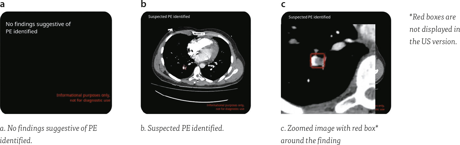 Figure 1. Automation Platform Pulmonary Embolism (CINA® PE): The Automation Platform Pulmonary Embolism has the possibility of a triage and notification of PE scans and flagging and communicating suspected or even absent findings in a PACS. In case of a suspected finding it will and show the key images of the findings. Marked in order for the radiologist to determine if immediate action is required.
Automation Platform Aortic Dissection (CINA® AD)
Aortic dissection is not common, and mostly presents as an acute condition in patients with catastrophic illness. However, early and accurate diagnosis and treatment is crucial for patient survival. CT is the most common modality of choice because of its widespread availability in emergency departments.
“The Automation Platform flags no findings of aortic dissection, or in the case of positive findings, it will indicate the presence and its location of the dissection. In the case of multiple dissections, it provides multiple key images so you can see the whole extent,” said Prof. Niehues. “Again, here, the median processing time is a little over 34 seconds, so it's very, very fast. And there is an even greater sensitivity of 96% and a specificity of 97%. You can really rely on the results being presented by the Automation Platform.”
Aortic dissection is not common, and mostly presents as an acute condition in patients with catastrophic illness. However, early and accurate diagnosis and treatment is crucial for patient survival. CT is the most common modality of choice because of its widespread availability in emergency departments.
“The Automation Platform flags no findings of aortic dissection, or in the case of positive findings, it will indicate the presence and its location of the dissection. In the case of multiple dissections, it provides multiple key images so you can see the whole extent,” said Prof. Niehues. “Again, here, the median processing time is a little over 34 seconds, so it's very, very fast. And there is an even greater sensitivity of 96% and a specificity of 97%. You can really rely on the results being presented by the Automation Platform.”
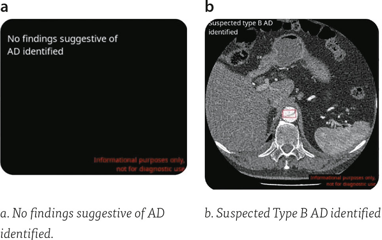 Figure 2. Automation Platform Aortic Dissection (CINA® AD): The Automation Platform Aortic Dissection (CINA® AD) shows thumbnails in PACS which mention no findings of aortic dissection, or in case of positive findings it will show the key image with a red box* to show the dissection. It will also classify the finding e.g. Type B aortic dissection.
Detecting these conditions is one thing, but presentations must be visible and understandable for physicians. “The Automation Platform has a web interface that highlights cases with positive findings. You have the opportunity to triage those patients, so you don't lose time to report on these. Some findings come with insights and results which will be or can be sent via email. And if you use push notification,even if you are not right in the emergency department, you get an email notification with the key images provided. So you do not lose any time for those patients with positive findings,” explained Professor Niehues.
“It's a zero-click solution, so it just works on its own. The results will be within your PACS in a median of 60 seconds.”
Combining the best of both worlds in Coronary CTA
Patients with coronary artery disease can benefit from use of Canon’s Super-Resolution Deep Learning Reconstruction (SL-DLR) technique, PIQE. Professor Mickaël Ohana, Consultant Radiologist at the Strasbourg University Hospital in France, explained how it provides clinicians with the possibility for higher diagnostic confidence and clarity as compared to conventional image reconstruction approaches in visualizing small arteries, plaques, and fine cardiac structures, and has the potential to assist clinicians in coronary atherosclerosis patients' cardiovascular risk stratification.
“PIQE directly brings the potential advantages of Ultra High Resolution CT (UHR-CT) to conventional CT. It is about merging the best of both worlds,” said Professor Ohana.
“Current research is focused on the advantages induced by very thin slice-thickness and increased matrix size. Mostly a sharper image quality, reduced artifacts, partial volume and blooming, and also an improved detection and characterization of small anatomical structures,” he continued.
“Through this, Super Resolution CT has shown promise in correctly identifying non-obstructive diseases that were labeled as obstructive with conventional CT.”
“We have noticed significant noise reduction, and increased sharpness of all the vascular and anatomical structures with PIQE compared to deep learning or hybrid IR reconstructions. The conspicuity of the structures and the contour can be more easily seen. Even in lesions with very subtle arterial calcification, PIQE provides a better view. This is the same with curved MPR - the sharpness of the vessels is better with PIQE. You can also achieve better delineation of smaller arterial branches. And you get an increased conspicuity of calcifications, mostly on faint or subtle low-density calcifications,” remarked Prof. Ohana. “All these points - the noise reduction, the increased sharpness, better delineation of small structures, and the increased conspicuity of calcifications - lead to a higher image quality with PIQE”.
“In the end, in routine clinical work, you can expect to get the advantages of the Super Resolution CT images, but without the drawbacks,” he added. “And at a lower cost with, of course, a higher number of machines to be able to do that. The availability of a wide area detector, which is something that once you get it, you cannot go back, when you do cardiac imaging. The faster rotation time, the ability to use systematically for all patients on 100kV and all that at the lowest radiation dose. This is really like combining best of both worlds.”
“What is important is that you can use it without any impact on the workflow or the dosimetry. You have an increased perceived image quality of coronary CTA, and in the end, it could carry a potential diagnostic impact as we have seen, probably a better assessment of highly calcified vessels, possibly also a better delineation of minimal atherosclerotic lesion, and also maybe a possible better plaque quantification,” he concluded. “It is very promising, and I think it's only the beginning of the technology. We hope that we will see more in the future.”
Better body imaging with MRI
Patient comfort, optimization of sequences, and improvement of image quality using new artificial intelligence technologies are key, especially in certain patient groups. They can lead to a better diagnosis and facilitate appropriate personalized treatment. Dr. Benoît Sauer, Radiologist at the Groupe d'imagerie médicale - MIM, Clinique Sainte Anne Strasbourg, in France, outlined the practical implications for improving body imaging of super-high resolution PIQE for MRI.
“Deep Learning Reconstruction is a major additional layer for improvement of image quality and also time in MRI, which is very important for patients,” said Dr. Sauer. “In addition, radiologists have more choice and more capacity to adapt to make better images for each patient characteristic.”
In abdominal imaging, quality is essential to make an accurate diagnosis. For upper abdomen scanning, the length of the patient’s breath-hold is a limit. For whole body diffusion scanning, the duration of the examination can be problematic for some patients. For whole-body DWI diffusion, for myeloma, comfort is very important. For musculoskeletal imaging, especially trauma-related scanning, total scanning time needs to be fast.
“It's a zero-click solution, so it just works on its own. The results will be within your PACS in a median of 60 seconds.”
Combining the best of both worlds in Coronary CTA
Patients with coronary artery disease can benefit from use of Canon’s Super-Resolution Deep Learning Reconstruction (SL-DLR) technique, PIQE. Professor Mickaël Ohana, Consultant Radiologist at the Strasbourg University Hospital in France, explained how it provides clinicians with the possibility for higher diagnostic confidence and clarity as compared to conventional image reconstruction approaches in visualizing small arteries, plaques, and fine cardiac structures, and has the potential to assist clinicians in coronary atherosclerosis patients' cardiovascular risk stratification.
“PIQE directly brings the potential advantages of Ultra High Resolution CT (UHR-CT) to conventional CT. It is about merging the best of both worlds,” said Professor Ohana.
“Current research is focused on the advantages induced by very thin slice-thickness and increased matrix size. Mostly a sharper image quality, reduced artifacts, partial volume and blooming, and also an improved detection and characterization of small anatomical structures,” he continued.
“Through this, Super Resolution CT has shown promise in correctly identifying non-obstructive diseases that were labeled as obstructive with conventional CT.”
“We have noticed significant noise reduction, and increased sharpness of all the vascular and anatomical structures with PIQE compared to deep learning or hybrid IR reconstructions. The conspicuity of the structures and the contour can be more easily seen. Even in lesions with very subtle arterial calcification, PIQE provides a better view. This is the same with curved MPR - the sharpness of the vessels is better with PIQE. You can also achieve better delineation of smaller arterial branches. And you get an increased conspicuity of calcifications, mostly on faint or subtle low-density calcifications,” remarked Prof. Ohana. “All these points - the noise reduction, the increased sharpness, better delineation of small structures, and the increased conspicuity of calcifications - lead to a higher image quality with PIQE”.
“In the end, in routine clinical work, you can expect to get the advantages of the Super Resolution CT images, but without the drawbacks,” he added. “And at a lower cost with, of course, a higher number of machines to be able to do that. The availability of a wide area detector, which is something that once you get it, you cannot go back, when you do cardiac imaging. The faster rotation time, the ability to use systematically for all patients on 100kV and all that at the lowest radiation dose. This is really like combining best of both worlds.”
“What is important is that you can use it without any impact on the workflow or the dosimetry. You have an increased perceived image quality of coronary CTA, and in the end, it could carry a potential diagnostic impact as we have seen, probably a better assessment of highly calcified vessels, possibly also a better delineation of minimal atherosclerotic lesion, and also maybe a possible better plaque quantification,” he concluded. “It is very promising, and I think it's only the beginning of the technology. We hope that we will see more in the future.”
Better body imaging with MRI
Patient comfort, optimization of sequences, and improvement of image quality using new artificial intelligence technologies are key, especially in certain patient groups. They can lead to a better diagnosis and facilitate appropriate personalized treatment. Dr. Benoît Sauer, Radiologist at the Groupe d'imagerie médicale - MIM, Clinique Sainte Anne Strasbourg, in France, outlined the practical implications for improving body imaging of super-high resolution PIQE for MRI.
“Deep Learning Reconstruction is a major additional layer for improvement of image quality and also time in MRI, which is very important for patients,” said Dr. Sauer. “In addition, radiologists have more choice and more capacity to adapt to make better images for each patient characteristic.”
In abdominal imaging, quality is essential to make an accurate diagnosis. For upper abdomen scanning, the length of the patient’s breath-hold is a limit. For whole body diffusion scanning, the duration of the examination can be problematic for some patients. For whole-body DWI diffusion, for myeloma, comfort is very important. For musculoskeletal imaging, especially trauma-related scanning, total scanning time needs to be fast.
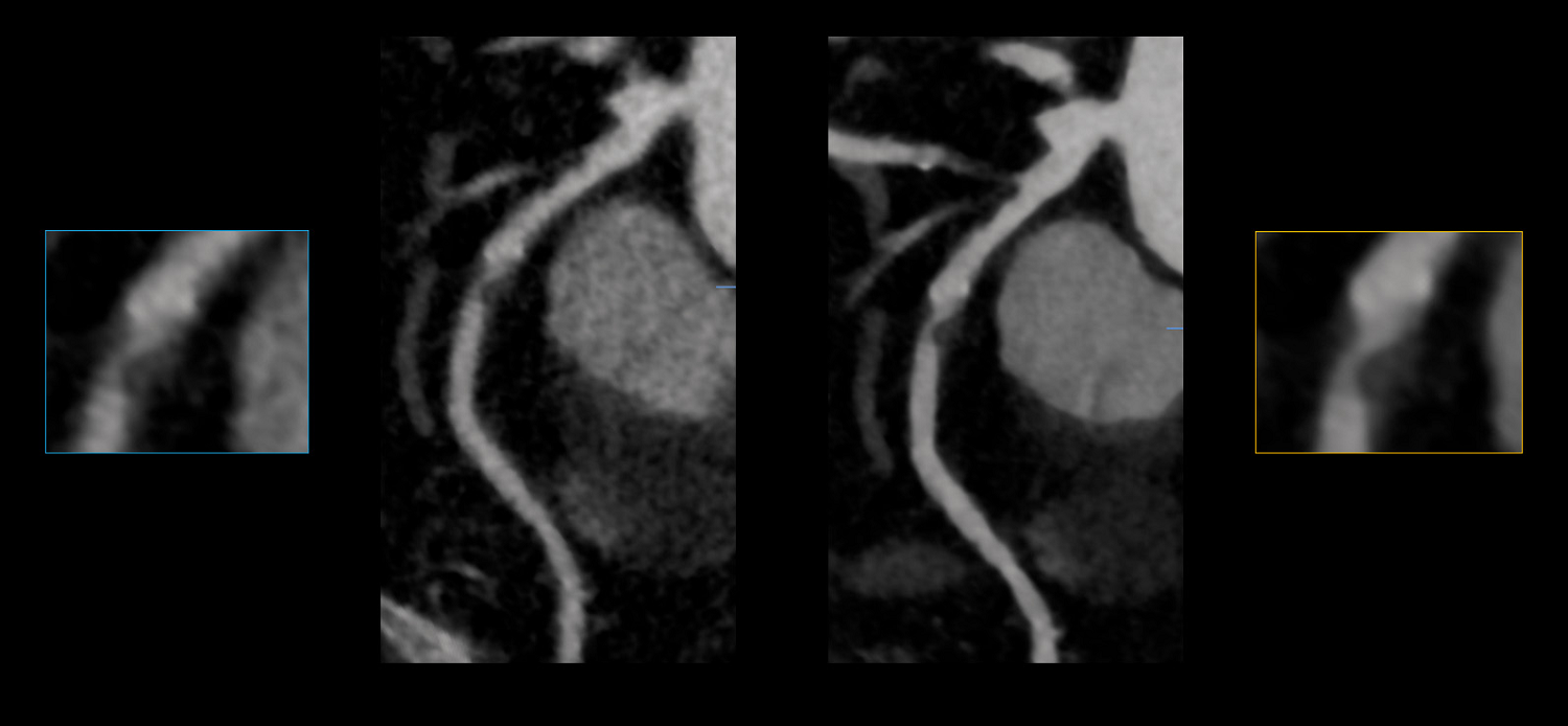 Figure 3. PIQE – CT Super Resolution-Deep Learning Reconstruction: The coronary artery CT image on the left (a) is reconstructed with Iterative Reconstruction and on the right (b) with Super Resolution-Deep Learning Reconstruction. The contour of the plaque can be better seen on the PIQE image and also a much better differentiation of the residual lumen inside can be seen compared to iterative reconstruction.
“With Canon’s Deep Learning Reconstruction techniques, AiCE and PIQE, we now have an MRI liver protocol of less than ten minutes, complete with a low dose CT scan of the whole thorax-abdomen. For unwell patients experiencing difficulties with breath-hold, we make shorter sequences of 13 seconds in dynamics, so we have high quality. For prostate, we can perform an examination in ten minutes,” said Dr. Sauer. “It works also for other things.
For example, musculoskeletal imaging and knee trauma with a rupture of LCA, and bone marrow oedema. The total examination is less than six minutes. The advantage is quicker results with fewer movement artifacts, so it's very important. We can obtain good quality images as well as a good diagnosis, fast.”
“In conclusion, quality and comfort in MRI are essential for the patient. With Deep Learning Reconstruction, our standard protocols now take less than 10 minutes in total to perform, except full body. AI allows for shorter examination times. It's more comfortable for the patient and improves the image quality. It also allows a personalized evaluation, rapid sequences to avoid movement artifacts and meets the needs of fragile patients,” he added. “These are major gains, especially in time and quality in MRI for the comfort of the patient.”
Meaningful innovation. Made possible.
Canon Medical is developing intelligent technologies to make a whole new level of quality, insight, and value across the entire care pathway possible. With these insights, it is becoming apparent that AI-based healthcare support can be successfully embedded into clinical practice for the benefit of patients and clinicians.
For example, musculoskeletal imaging and knee trauma with a rupture of LCA, and bone marrow oedema. The total examination is less than six minutes. The advantage is quicker results with fewer movement artifacts, so it's very important. We can obtain good quality images as well as a good diagnosis, fast.”
“In conclusion, quality and comfort in MRI are essential for the patient. With Deep Learning Reconstruction, our standard protocols now take less than 10 minutes in total to perform, except full body. AI allows for shorter examination times. It's more comfortable for the patient and improves the image quality. It also allows a personalized evaluation, rapid sequences to avoid movement artifacts and meets the needs of fragile patients,” he added. “These are major gains, especially in time and quality in MRI for the comfort of the patient.”
Meaningful innovation. Made possible.
Canon Medical is developing intelligent technologies to make a whole new level of quality, insight, and value across the entire care pathway possible. With these insights, it is becoming apparent that AI-based healthcare support can be successfully embedded into clinical practice for the benefit of patients and clinicians.
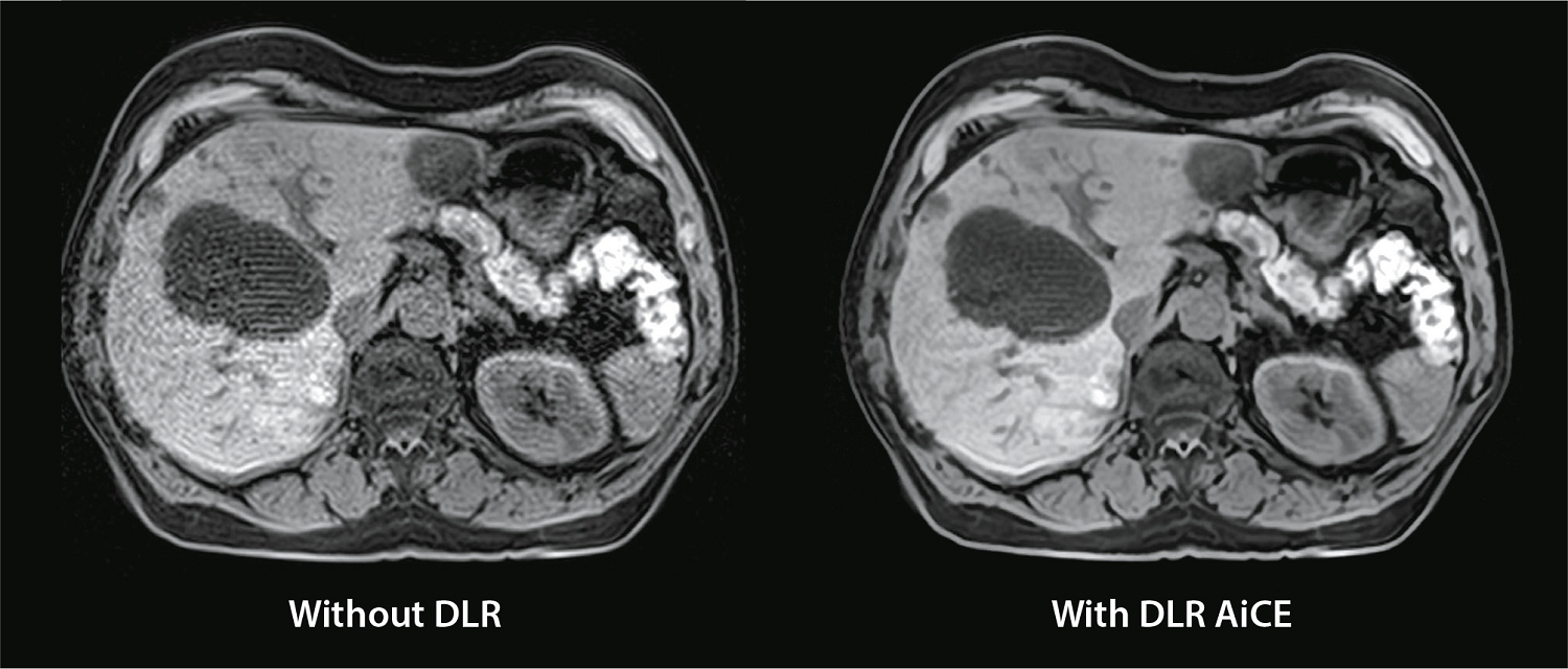 Figure 4. MRI Deep Learning Reconstruction: For whole body MRI diffusion the duration of the examination could be problematic. An MRI diffusion image of the abdomen of 15 sec is reconstructed without DLR on the left (a) and with DLR (AiCE) (b) on the right. There is a great increase of image quality in the DLR image. In MRI, shorter scan time equals to quality and comfort for the patient.

Prof. Stefan M. Niehues, MD, MHBA
Head physician of the Department of Radiology,
Caritas Clinics Berlin, Germany

Prof. Mickaël Ohana, MD, PhD
Consultant Radiologist
Strasbourg University
Hospital Strasbourg, France.

Dr. Benoît Sauer, MD
Radiologist Groupe d'imagerie médicale - MIM,
Clinique Sainte Anne Strasbourg, France.
Watch the full webinar here.
Other blogs that may interest you
Disclaimers
Social Media


