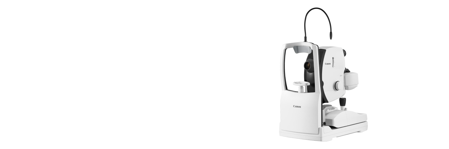
The multifaceted CX-1

The CX-1 is a Mydriatic Retinal Camera that can be changed into a Non-Mydriatic camera by a simple push of a button.
Besides color photography, the CX-1 can perform FLUO, Red Free, Cobalt and standard even FAF photography.

All photography modes can be performed in either the MYD or NON MYD mode. This provides exceptional versatility and enables diagnosis, screening and monitoring of all major eye diseases.

sou
Color
Establishing base Line
sou
Fluo
Checking retinal flow for occlusions and leakage

sou
FAF
Can contribute to the early detection of retinal changes
sou
Red Free
Enhances the visibility of retinal vasculature, hemorrhages, drusen and exudates
sou
Cobalt
Provides a dark background that enhances the visualization of the Nerve Fiber Layer

RGB channels

In addition to the optical filters of the CX-1 , the RX software offers also the possibility of a RGB channel filter. The Red channel can be helpful for the visualization of pigmentary disturbances, choroidal ruptures, nevi and malignant melanomas can contribute to the early detection of retinal changes.

Unrivalled Image quality

Superb Canon optics and a dedicated 24 MP digital camera for highest image quality.
Carefully selected optical filter sets provide highest image quality for Fluorescein angiography and FAF.

Canon Opacity suppression will largely suppresses the effects of cataracts and other ocular opacities in color images.

Easy Operation

The CX-1 is a compact device that allows for maximal patient interaction. It requires only a short reaching distance for easy opening patient’s eyelid.
All filters are motorized; as is the chinrest adjustment- easy to operate by a push of a button!

Panning and tilting

The standard panning and tilting feature enables to work around central ocular obstructions (cataracts, vitreous hemorrhages. And it can assist in imaging the peripheral retina.
Tilting operation

Automatically optimized flash range

The CX-1 has an automatically optimized flash range, -adjusted to the different photography modes and ISO settings. It will help you by automatically selecting one of the 168 possible flash settings.

Non Mydriatic Mode

The Non-Mydriatic mode is essential for non dilatable patients such as glaucoma suspects. Children and photosensitive patients will also benefit from the non invasive IRED observation light.
In NM mode the observation is done on the observation screen of the digital camera and while using invisibIe IR observation light.
The imaging angle in this mode is 45 degrees (or 67.5 degrees in central angle of view annotation).

Color, Red-free, Cobalt, Fundus Autofluorescence (FAF) and even Fluorescein Angiography can be performed in this mode.

sou
sou

Mydriatic Mode

In this more traditional operation, the observation is done with viewfinder, while using visible observation light. It will provide a clear view of the retina in color The imaging angle in this mode is 50 degrees (or 75 degrees, central angle of view).


sou
sou

Mosaic

Use the mosaic function in the RX software to create one high resolution wide field image (up to 85 degrees).
Up to 20 individual images can be combined.

2X magnification

In addition the CX-1 is equipped with a 2X button that provides a 2x digital magnification or depending on a setting, a 30 degrees image.

30 degrees angle 

A 30 degrees angle is required for participation in studies and co-operation with most reading centers: i.e DARC, Wisconsin and VRC.

Fundus Autofluorescence (FAF)

A diagnostic technique for documenting the deposition of lipofuscin in the retinal pigment epithelium (RPE). It is easy and non invasive and provides information that may otherwise not be clinically detectable.
It can be a valuable asset in diagnosing retinal disease.

Since the introduction of fluorescein angiography (FFA) in 1959, ophthalmologists observed that even without the use of fluorescein, parts of the fundus would show areas of faint fluorescence under certain conditions. This naturally occurring fluorescence is mainly caused by Lipofuscin. Lipofuscin is a fluorescent pigment that accumulates in the RPE as a metabolic byproduct of cell function.
Lipofuscin deposition normally increases with age, but may also occur from RPE cell dysfunction or an abnormal metabolic load on the RPE. FAF imaging can visualize the deposition of lipofuscin in the retinal pigment epithelium (RPE). Areas of excess Lipofuscin accumulation will appear hyperfluorescent. But when RPE cells die or are absent, LF disappears, leading to hypofluorescence.

Green Spectrum

The CX-1 utilizes green light spectrum to stimulate the retina and captures the emission in the yellow-orange spectrum- utilizing carefully selected matching optical filters. Using the green spectrum may provide more detail in the fovea, as where blue light tends to be absorbed by the high concentration of macular pigments. Additional advantage of green light is that the longer wavelength tends to have less absorption by the crystalline lens of the eye, especially in patients with cataracts.

Am I missing something without FAF?

In this example it is clear that the FAF image is showing additional clinical information that can’t be seen in the color image!

sou
sou

Karolinska Instutet

“With the extra feature of FAF photography we have discovered retinal changes we have not seen before and which makes us earn more about retinal changes and diseases every day“

Rune Brautaset
Professor
Vice-chairman Committee for Higher Education
Head of division for Eye and Vision
Karolinska Institutet, Stockholm
Contact Us