
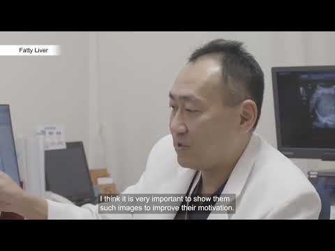
Attenuation Imaging (ATI)
Quantify and color-code changes in the liver composition (e.g. fat content)
Using statistical analysis of the attenuation throughout the region of interest, the system is able to quantify the accuracy of the sample. This provides information regarding the uniformity of the attenuation which is important for accuracy.
- As the ultrasound wave is transmitted though tissue, it is attenuated through the process of scatter, absorption and heat.
- The amount of attenuation is proportional to the frequency.
Shear Wave Elastography (SWE)
Non-invasive, quantitative assessment of tissue stiffness
- Overcoming complications / limitations of biopsy
- 5 smart maps to visualise and quantify shear wave propagation in real time, including a variance map
- Canon unique propagation maps actually show shearwave propagation through the tissue
- This assists in measurement placement ensuring accuracy, reproducibility and best practice
- One shot or continuous mode scanning allowing multiple measurements from one acquisition and quicker examination times
- Push pulse optimised for deeper regions
Shear waves are generated by means of an ultrasonic burst (left). Depending on tissue properties, shear waves travel at varying speed. Canon's unique propagation mode can be used to confirm the quality of the shear wave generation (right).
Transducer technology
Wideband multi-frequency iDMS convex: i8CX1 (PVI-475BX)
Aplio i-series provides superior sensitivity and resolution for both near and far field. The ability to use one transducer across a wider range of patient types can potentially reduce cost, while providing better imaging.
Features:
- Up to 50 cm penetration

Multi-frequency slim-face convex: i8C1 (PVI-475BT)
This narrow face transducer features:
- Improved shape for easy grip
- Super lightweight transducer head
- Light weight, thin and highly flexible cable

Multi-frequency micro-convex: 8MC1 (PVT-482BT)
Aplio i-series' thin micro-convex provides enhanced maneuverability and ease of access.
New Features:
- Single crystal technology
- Lightweight and maneuverable
- New CIVCO® Verza™ Guidance System
Clinical Applications:
- Abdominal
- Pediatric
- OB
- Biopsy Guidance

Laparoscopic liver resection as a less invasive treatment option for hepatocellular carcinoma
In this white paper, Dr. Satoru Seo, Department of Surgery from the Graduate School of Medicine, Kyoto University, shared his clinical impressions of the PET-805LA transducer used in combination with the Aplio series diagnostic ultrasound system and discussed the clinical usefulness of intraoperative contrast-enhanced ultrasound.
Education & resources


Available Systems
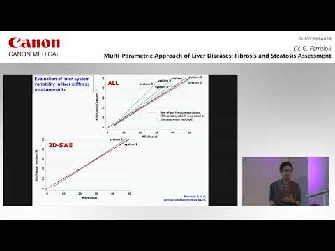
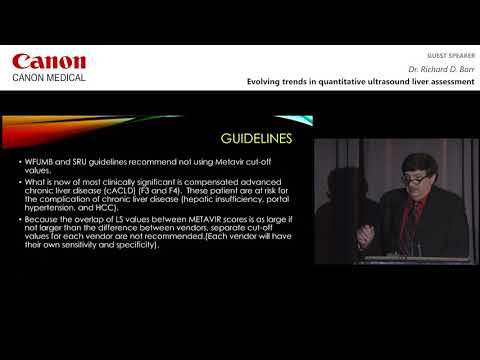
New WFUMB guidelines - Liver Ultrasound Elastography: An Update to the World Federation for Ultrasound in Medicine and Biology Guidelines and Recommendations. (Page 2422 table 2)
SWE
SWE acquisition. Propagation lines in the far field represent how the shear wave has been affected by the vessels in the ROI and loss of data is shown here as lack of colour fill in the speed map. This indicates an area where calliper placement should not be performed but rather in areas of homogeneity on the colour map and continuous parallel propagation lines
SWE using dual mode together with speed and propagation maps. We use both maps to help with accuracy and reproducibility in our calliper placement, extremely important when undertaking SWE. We can use the speed map (or an elastography map), to look for uniformity in data collection, areas of black indicate no data acquired and callipers should not be placed here. Propagation maps are used to look at continuous parallel lines to also indicate good quality data and correct calliper placement.
A case of liver fibrosis. Different colours are seen within the speed map indicating areas of increased stiffness. Propagation lines are also thicker and further apart – another indication of increased stiffness. Both correlate to an increase in number, suggestive of fibrosis.
New WUFUMB guidelines* recommend only one measurement be taken from each Shear Wave acquisition. With continuous wave acquisition, the Canon systems can acquire a ‘cine loop’ of Shear Waves to make multiple measurements quicker and easier.
ATI
A good acquisition using ATI. Values are shown in white representing a good quality acquisition.
A good acquisition using ATI. Values are shown in white representing a good quality acquisition.
A good acquisition using ATI. Values are shown in white representing a good quality acquisition.
Another ATI acquisition, this time showing values in yellow representing a mediocre acquisition.
ATI showing values in red representing a poor acquisition
SWE QuadView
Social Media


