


Satellite Symposia at ECR 2026
Building on the strong participation in recent years, Canon Medical Academy has doubled its offering for the European Congress of Radiology (ECR) 2026 in Vienna, with two dedicated satellite symposia supporting our continuous education program.
Together, these sessions aim to help push the boundaries of knowledge in everyday radiology practice.
ECR 2026 | Satellite Symposium | Canon Medical Academy Advancing CEUS in Radiology: Clinical Innovations, Faster Pathways, Higher Efficiency | Symposium
Thursday, March 5, 2026, 14:30–15:30 (60 min) | Room G2

ECR 2026 | Satellite Symposium | Canon Medical Academy The Next Level of Diagnostic Quality in Photon Counting and Area Detector CT: AI, Spectral, and UHR | Symposium
Friday, March 6, 2026, 09:30–10:30 (60min), Room N

Moderator: Prof. Adrian Lim
Professor and Consultant Radiologist
Imperial College London and the Healthcare NHS Trust
London, United Kingdom
Professor and Consultant Radiologist
Imperial College London and the Healthcare NHS Trust
London, United Kingdom

Speaker: Prof. Thomas Fischer
Radiologist, Head of Interdisciplinary Ultrasound Center,
Radiology, Executive Senior Physician, Department of Radiology
Charité University Medicine
Berlin, Germany
Presentation: State of the Art Abdominal CEUS Clinical Innovations
Abstract: Explore the clinical applications of contrast-enhanced ultrasound (CEUS) in radiology and review the latest innovations and supporting evidence shaping its use in patient care.
Radiologist, Head of Interdisciplinary Ultrasound Center,
Radiology, Executive Senior Physician, Department of Radiology
Charité University Medicine
Berlin, Germany
Presentation: State of the Art Abdominal CEUS Clinical Innovations
Abstract: Explore the clinical applications of contrast-enhanced ultrasound (CEUS) in radiology and review the latest innovations and supporting evidence shaping its use in patient care.

Speaker: Prof. Maija Radzina
Tenured Professor
Pauls Stradiņš Clinical University Hospital
Riga, Latvia
European Radiology Society, Executive Council member
Presentation: Diagnostic and Economic Benefits of CEUS in Clinical Routine
Abstract: Discover how contrast-enhanced ultrasound (CEUS) streamlines patient pathways, improves diagnostic efficiency, and enhances modern radiology practice by integrating its benefits into routine imaging.
Tenured Professor
Pauls Stradiņš Clinical University Hospital
Riga, Latvia
European Radiology Society, Executive Council member
Presentation: Diagnostic and Economic Benefits of CEUS in Clinical Routine
Abstract: Discover how contrast-enhanced ultrasound (CEUS) streamlines patient pathways, improves diagnostic efficiency, and enhances modern radiology practice by integrating its benefits into routine imaging.

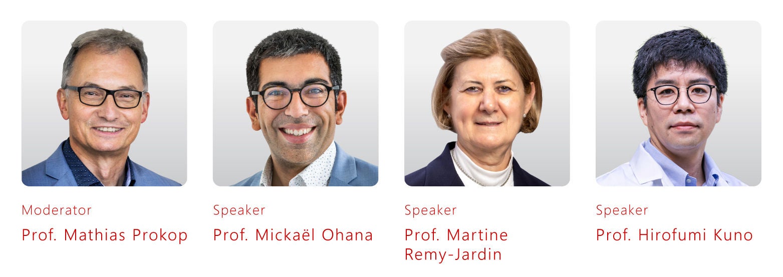
Moderator: Prof. Mathias Prokop
Radiologist, Professor, and Chairman, Department of Medical Imaging
Radboud University Medical Center
Nijmegen, the Netherlands
Radiologist, Professor, and Chairman, Department of Medical Imaging
Radboud University Medical Center
Nijmegen, the Netherlands

Speaker: Prof. Mickaël Ohana
Consultant Radiologist
Strasbourg University Hospital
Strasbourg, FRANCE
Presentation: Latest Deep Learning Technologies in Cardiac CT: One-Beat Super-Resolution Imaging with Whole-Heart Motion Correction
Abstract: Achieving optimal image quality in cardiac CT imaging requires a combined high spatial and temporal resolution to minimize blooming and motion artifacts. This presentation will show how this can be achieved with wide-area detector CT coupled with the latest deep learning technologies. Thanks to a fast gantry rotation time and the application of deep learning-based motion correction (CLEAR Motion Cardiac), new levels of temporal resolution can be achieved, enabling the acquisition in a single heartbeat even in patients with high heart rates.
Furthermore, super-resolution deep learning reconstruction (PIQE) provides images at improved spatial resolution, going beyond the size of the physical detector elements. As a result of these technologies, cardiac CT imaging has become feasible and accessible to all patients, including those with high calcium scores, high heart rates, and high body mass index. In this presentation, these technologies will be explained, and both clinical cases and the latest scientific findings will be presented.
Consultant Radiologist
Strasbourg University Hospital
Strasbourg, FRANCE
Presentation: Latest Deep Learning Technologies in Cardiac CT: One-Beat Super-Resolution Imaging with Whole-Heart Motion Correction
Abstract: Achieving optimal image quality in cardiac CT imaging requires a combined high spatial and temporal resolution to minimize blooming and motion artifacts. This presentation will show how this can be achieved with wide-area detector CT coupled with the latest deep learning technologies. Thanks to a fast gantry rotation time and the application of deep learning-based motion correction (CLEAR Motion Cardiac), new levels of temporal resolution can be achieved, enabling the acquisition in a single heartbeat even in patients with high heart rates.
Furthermore, super-resolution deep learning reconstruction (PIQE) provides images at improved spatial resolution, going beyond the size of the physical detector elements. As a result of these technologies, cardiac CT imaging has become feasible and accessible to all patients, including those with high calcium scores, high heart rates, and high body mass index. In this presentation, these technologies will be explained, and both clinical cases and the latest scientific findings will be presented.
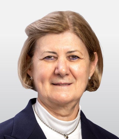
Speaker: Prof. Martine Remy-Jardin
Chest Radiologist
Radboud University Medical Center
Nijmegen, the Netherlands
Presentation: Photon-Counting CT in Chest Imaging: Most Promising Applications to Improve Patient Management
Abstract: It is now clear that photon-counting CT fulfills the expectations of improved spatial resolution and dose reduction, and holds the promise of improving patient management. In this session, the most recent results on photon-counting CT in chest imaging will be presented, with a focus on a cadmium-zinc-telluride (CZT)-based system equipped with deep-learning reconstruction technology. In parallel to the description of this technology and its potential, early clinical findings will be presented, and the potential to improve patient management will be discussed.
Chest Radiologist
Radboud University Medical Center
Nijmegen, the Netherlands
Presentation: Photon-Counting CT in Chest Imaging: Most Promising Applications to Improve Patient Management
Abstract: It is now clear that photon-counting CT fulfills the expectations of improved spatial resolution and dose reduction, and holds the promise of improving patient management. In this session, the most recent results on photon-counting CT in chest imaging will be presented, with a focus on a cadmium-zinc-telluride (CZT)-based system equipped with deep-learning reconstruction technology. In parallel to the description of this technology and its potential, early clinical findings will be presented, and the potential to improve patient management will be discussed.

Speaker: Prof. Hirofumi Kuno
Section Head, Department of Diagnostic Radiology
National Cancer Center Hospital East
Kashiwa, Japan
Presentation: Clinical Applications of Photon-Counting CT in Head and Neck Cancer imaging
Abstract: CT imaging for head and neck cancer has advanced with dual-energy CT and ultra-high-resolution systems such as the Aquilion Precision CT, enhanced by deep-learning reconstruction (AiCE) for improved image quality at reduced dose. Photon-counting CT (PCCT) represents a further development, offering high spatial resolution and true multi-energy spectral data in a single acquisition. This session will discuss practical PCCT protocols for head and neck imaging and demonstrate how these capabilities enhance diagnostic performance in routine clinical practice. The integration of deep-learning–based noise reduction in PCCT and its clinical impact will also be discussed.
Section Head, Department of Diagnostic Radiology
National Cancer Center Hospital East
Kashiwa, Japan
Presentation: Clinical Applications of Photon-Counting CT in Head and Neck Cancer imaging
Abstract: CT imaging for head and neck cancer has advanced with dual-energy CT and ultra-high-resolution systems such as the Aquilion Precision CT, enhanced by deep-learning reconstruction (AiCE) for improved image quality at reduced dose. Photon-counting CT (PCCT) represents a further development, offering high spatial resolution and true multi-energy spectral data in a single acquisition. This session will discuss practical PCCT protocols for head and neck imaging and demonstrate how these capabilities enhance diagnostic performance in routine clinical practice. The integration of deep-learning–based noise reduction in PCCT and its clinical impact will also be discussed.
Moderator: Prof. Adrian Lim
Professor and Consultant Radiologist
Imperial College London and the Healthcare NHS Trust
London, United Kingdom
Speaker: Prof. Thomas Fischer
Radiologist, Head of Interdisciplinary Ultrasound Center,
Radiology, Executive Senior Physician, Department of Radiology
Charité University Medicine
Berlin, Germany
Presentation: State of the Art Abdominal CEUS Clinical Innovations
Abstract: Explore the clinical applications of contrast-enhanced ultrasound (CEUS) in radiology and review the latest innovations and supporting evidence shaping its use in patient care.
Speaker: Prof. Maija Radzina
Tenured Professor
Pauls Stradiņš Clinical University Hospital
Riga, Latvia
European Radiology Society, Executive Council member
Presentation: Diagnostic and Economic Benefits of CEUS in Clinical Routine
Abstract: Discover how contrast-enhanced ultrasound (CEUS) streamlines patient pathways, improves diagnostic efficiency, and enhances modern radiology practice by integrating its benefits into routine imaging.
Moderator: Prof. Mathias Prokop
Radiologist, Professor, and Chairman, Department of Medical Imaging
Radboud University Medical Center
Nijmegen, the Netherlands
Speaker: Prof. Mickaël Ohana
Consultant Radiologist
Strasbourg University Hospital
Strasbourg, FRANCE
Presentation: Latest Deep Learning Technologies in Cardiac CT: One-Beat Super-Resolution Imaging with Whole-Heart Motion Correction
Abstract: Achieving optimal image quality in cardiac CT imaging requires a combined high spatial and temporal resolution to minimize blooming and motion artifacts. This presentation will show how this can be achieved with wide-area detector CT coupled with the latest deep learning technologies. Thanks to a fast gantry rotation time and the application of deep learning-based motion correction (CLEAR Motion Cardiac), new levels of temporal resolution can be achieved, enabling the acquisition in a single heartbeat even in patients with high heart rates.
Furthermore, super-resolution deep learning reconstruction (PIQE) provides images at improved spatial resolution, going beyond the size of the physical detector elements. As a result of these technologies, cardiac CT imaging has become feasible and accessible to all patients, including those with high calcium scores, high heart rates, and high body mass index. In this presentation, these technologies will be explained, and both clinical cases and the latest scientific findings will be presented.
Speaker: Prof. Martine Remy-Jardin
Chest Radiologist
Radboud University Medical Center
Nijmegen, the Netherlands
Presentation: Photon-Counting CT in Chest Imaging: Most Promising Applications to Improve Patient Management
Abstract: It is now clear that photon-counting CT fulfills the expectations of improved spatial resolution and dose reduction, and holds the promise of improving patient management. In this session, the most recent results on photon-counting CT in chest imaging will be presented, with a focus on a cadmium-zinc-telluride (CZT)-based system equipped with deep-learning reconstruction technology. In parallel to the description of this technology and its potential, early clinical findings will be presented, and the potential to improve patient management will be discussed.
Speaker: Prof. Hirofumi Kuno
Section Head, Department of Diagnostic Radiology
National Cancer Center Hospital East
Kashiwa, Japan
Presentation: Clinical Applications of Photon-Counting CT in Head and Neck Cancer imaging
Abstract: CT imaging for head and neck cancer has advanced with dual-energy CT and ultra-high-resolution systems such as the Aquilion Precision CT, enhanced by deep-learning reconstruction (AiCE) for improved image quality at reduced dose. Photon-counting CT (PCCT) represents a further development, offering high spatial resolution and true multi-energy spectral data in a single acquisition. This session will discuss practical PCCT protocols for head and neck imaging and demonstrate how these capabilities enhance diagnostic performance in routine clinical practice. The integration of deep-learning–based noise reduction in PCCT and its clinical impact will also be discussed.
Disclaimers
- The opinions expressed in this material are solely those of the presenter and not necessarily those of Canon Medical Systems. Canon Medical Systems does not guarantee the accuracy or reliability of the information provided herein.
- The clinical results, performance and views described in this video/webinar are the experience of the clinician. Results may vary due to clinical setting, patient presentation and other factors. Many factors could cause the actual results and performance of Canon Medical’s products to be materially different from any of the aforementioned.
- Some products shown might not be available in all regulatory jurisdictions, please consult with your local Canon sales office for availability in your region.
Social Media


